

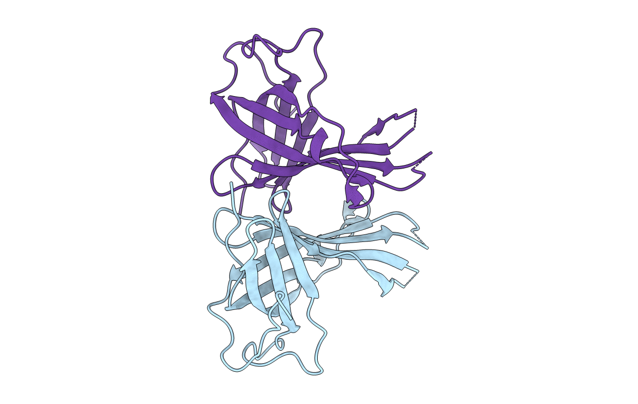
5XLM image
Deposition Date 2017-05-10
Release Date 2018-05-16
Last Version Date 2024-11-13
Entry Detail
PDB ID:
5XLM
Keywords:
Title:
Monomer form of M.tuberculosis PknI sensor domain
Biological Source:
Expression System(s):
Method Details:
Experimental Method:
Resolution:
2.20 Å
R-Value Free:
0.25
R-Value Work:
0.22
R-Value Observed:
0.22
Space Group:
P 31

Macromolecular Entities
Structures with similar UniProt ID
Protein Blast
Polymer Type:polypeptide(L)
Molecule:Serine/threonine-protein kina
Gene (Uniprot):pknI
Chain IDs:A, B
Chain Length:214
Number of Molecules:2
Biological Source:Mycobacterium tuberculosis (strain ATCC 25618 / H37Rv)
Primary Citation
Structural Insight into the Activation of PknI Kinase from M. tuberculosis via Dimerization of the Extracellular Sensor Domain.
Structure 25 1286 1294.e4 (2017)
PMID: 28712808 DOI: 10.1016/j.str.2017.06.010

Abstact

Protein kinases play central roles in the survival of Mycobacterium tuberculosis within host. Here we report the individual high-resolution crystal structures of the sensor domain (in both monomer and dimer forms) and the kinase domain of PknI, a transmembrane protein member of the serine/threonine protein kinases (STPKs) family. PknI is the first STPK identified whose sensor domain exists in a monomer-dimer equilibrium. Inspection of the two structures of the sensor domain (PknI_SD) revealed conformational changes upon dimerization, with an arm region of critical importance for dimer formation identified. Rapamycin-induced dimerization of unphosphorylated fusions of PknI juxtamembrane and the kinase domain, intended to mimic the dimerization effect presumably imposed by PknI_SD, was observed to be able to activate auto-phosphorylation activity of the kinase domain. In vivo experiments using an M. bovis model suggested PknI functions as a dimer in the regulation of M. tuberculosis growth.

Legend

Protein

Chemical

Disease

Primary Citation of related structures
Feedback Form
Name
Email
Institute
Feedback