Deposition Date
2017-03-30
Release Date
2018-02-07
Last Version Date
2023-11-22
Entry Detail
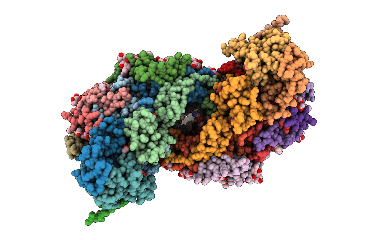
PDB ID:
5XDX
Keywords:
Title:
Bovine heart cytochrome c oxidase in the reduced state with pH 7.3 at 1.99 angstrom resolution
Biological Source:
Source Organism(s):
Bos taurus (Taxon ID: 9913)
Method Details:
Experimental Method:
Resolution:
1.99 Å
R-Value Free:
0.20
R-Value Work:
0.18
R-Value Observed:
0.18
Space Group:
P 21 21 21