Deposition Date
2016-12-02
Release Date
2017-03-22
Last Version Date
2024-10-30
Entry Detail
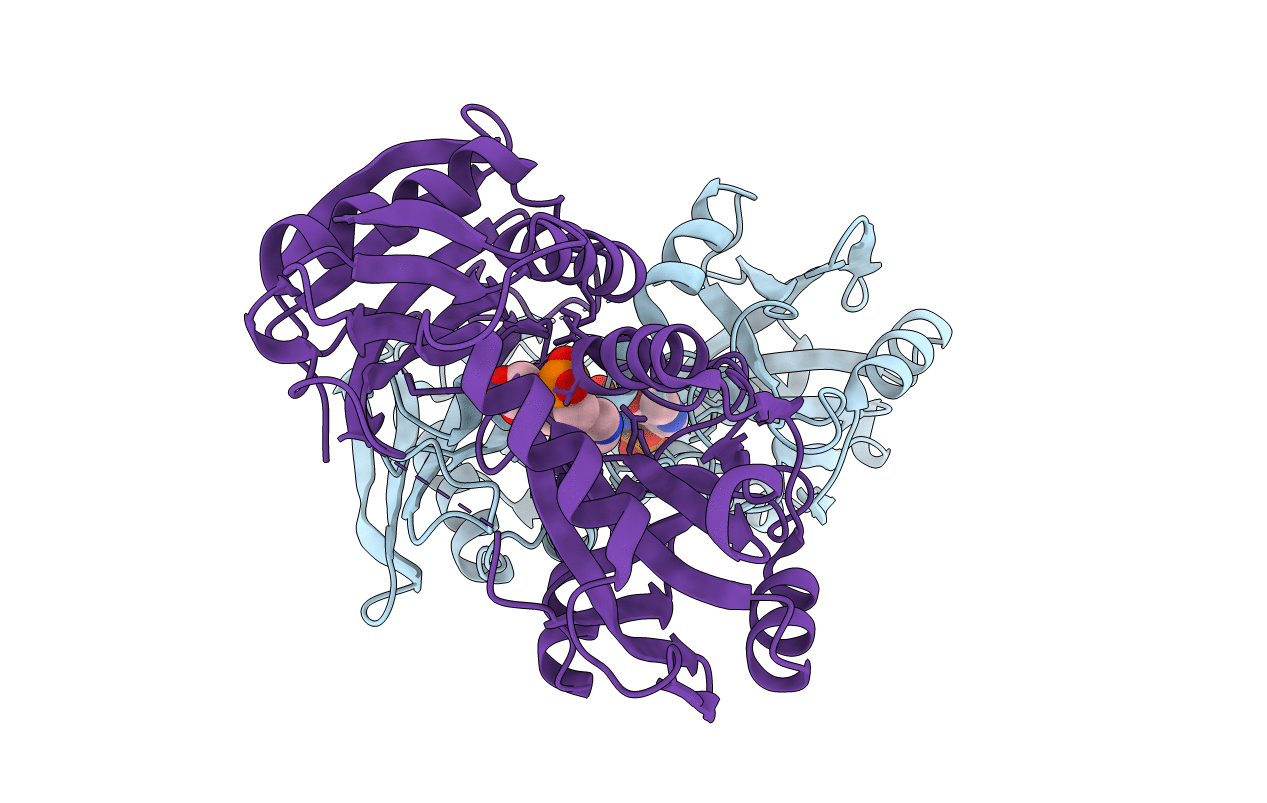
PDB ID:
5U3F
Keywords:
Title:
Structure of Mycobacterium tuberculosis IlvE, a branched-chain amino acid transaminase, in complex with D-cycloserine derivative
Biological Source:
Source Organism(s):
Expression System(s):
Method Details:
Experimental Method:
Resolution:
1.70 Å
R-Value Free:
0.20
R-Value Work:
0.17
R-Value Observed:
0.17
Space Group:
P 21 21 21