

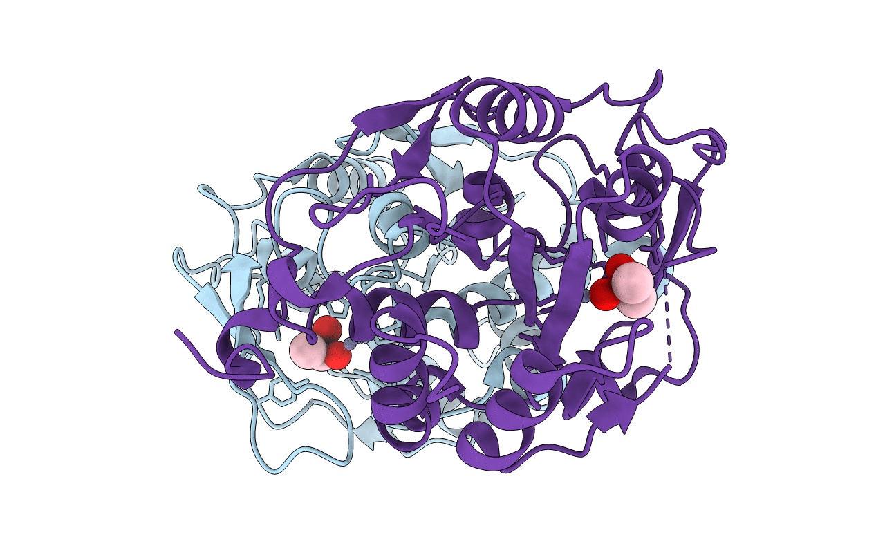
5HXD image
Deposition Date 2016-01-30
Release Date 2017-02-08
Last Version Date 2024-11-20
Entry Detail
PDB ID:
5HXD
Keywords:
Title:
Crystal structure of murein-tripeptide amidase MpaA from Escherichia coli O157
Biological Source:
Source Organism(s):
Expression System(s):
Method Details:
Experimental Method:
Resolution:
2.60 Å
R-Value Free:
0.24
R-Value Work:
0.17
R-Value Observed:
0.17
Space Group:
P 31

Macromolecular Entities
Structures with similar UniProt ID
Protein Blast
Polymer Type:polypeptide(L)
Molecule:Protein MpaA
Gene (Uniprot):mpaA
Chain IDs:A, B
Chain Length:237
Number of Molecules:2
Biological Source:Escherichia coli O157:H7
Primary Citation
Crystal Structure of Murein-Tripeptide Amidase MpaA from Escherichia coli O157 at 2.6 angstrom Resolution
Protein Pept.Lett. 24 181 187 (2017)
PMID: 27894248 DOI: 10.2174/0929866523666161128153128

Abstact

Peptidoglycan (PG) is an essential component of the cell wall, and undergoes reconstruction by various PG hydrolases during cell growth, development and division. The murein- tripeptide (Mtp) amidase MpaA belongs to PG hydrolase family and is responsible for cleaving the γ-D-Glumeso- Dap amide bond in the Mtp released during PG turnover. The current paper reports the crystal structure of MpaA from Escherichia coli (E. coli) O157 at 2.6 Å resolution. The asymmetric unit consists of two protein molecules and each monomer represents the common α/β fold of metallocarboxypeptidases (MCP). The Tyr133-Asp143 loop appears to mediate the entrance and binding of the substrate into the active groove. A structural comparison of MpaA with its homologue from Vibrio harveyi showed that MpaA has narrower active pocket entrance with a smaller surface opening, which is determined by the Val204-Thr211 loop. The reported structure provides a starting point for the molecular mechanism of MpaA in a significant human pathogen.

Legend

Protein

Chemical

Disease

Primary Citation of related structures
Feedback Form
Name
Email
Institute
Feedback