

5EW6 image
Deposition Date 2015-11-20
Release Date 2016-08-10
Last Version Date 2024-11-20
Entry Detail
PDB ID:
5EW6
Title:
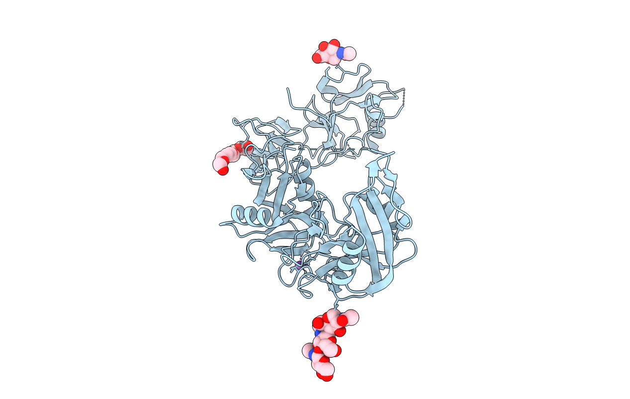
Structure of ligand binding region of uPARAP at pH 7.4 without calcium
Biological Source:
Source Organism(s):
Homo sapiens (Taxon ID: 9606)
Expression System(s):
Method Details:
Experimental Method:
Resolution:
2.29 Å
R-Value Free:
0.23
R-Value Work:
0.19
R-Value Observed:
0.19
Space Group:
P 41 21 2
Macromolecular Entities
Structures with similar UniProt ID
Protein Blast
Polymer Type:polypeptide(L)
Molecule:C-type mannose receptor 2
Gene (Uniprot):MRC2
Chain IDs:A
Chain Length:492
Number of Molecules:1
Biological Source:Homo sapiens
Peptide-like Molecules
PRD_900017
Primary Citation
Crystal structures of the ligand-binding region of uPARAP: effect of calcium ion binding
Biochem. J. 473 2359 2368 (2016)
PMID: 27247422 DOI: 10.1042/BCJ20160276

Abstact

The proteins of the mannose receptor (MR) family share a common domain organization and have a broad range of biological functions. Urokinase plasminogen activator receptor-associated protein (uPARAP) (or Endo180) is a member of this family and plays an important role in extracellular matrix remodelling through interaction with its ligands, including collagens and urokinase plasminogen activator receptor (uPAR). We report the crystal structures of the first four domains of uPARAP (also named the ligand-binding region, LBR) at pH 7.4 in Ca(2+)-bound and Ca(2+)-free forms. The first domain (cysteine-rich or CysR domain) folds into a new and unique conformation different from the β-trefoil fold of typical CysR domains. The so-called long loop regions (LLRs) of the C-type lectin-like domain (CTLD) 1 and 2 (the third and fourth domain) mediate the direct contacts between these domains. These LLRs undergo a Ca(2+)-dependent conformational change, and this is likely to be the key structural determinant affecting the overall conformation of uPARAP. Our results provide a molecular mechanism to support the structural flexibility of uPARAP, and shed light on the structural flexibility of other members of the MR family.

Legend

Protein

Chemical

Disease

Primary Citation of related structures
Feedback Form
Name
Email
Institute
Feedback