5C32 image
Deposition Date 2015-06-16
Release Date 2016-12-21
Last Version Date 2024-11-20
Entry Detail
PDB ID:
5C32
Title:
Constitutively active Sin recombinase cataltyic domain - I100T
Biological Source:
Source Organism(s):
Expression System(s):
Method Details:
Experimental Method:
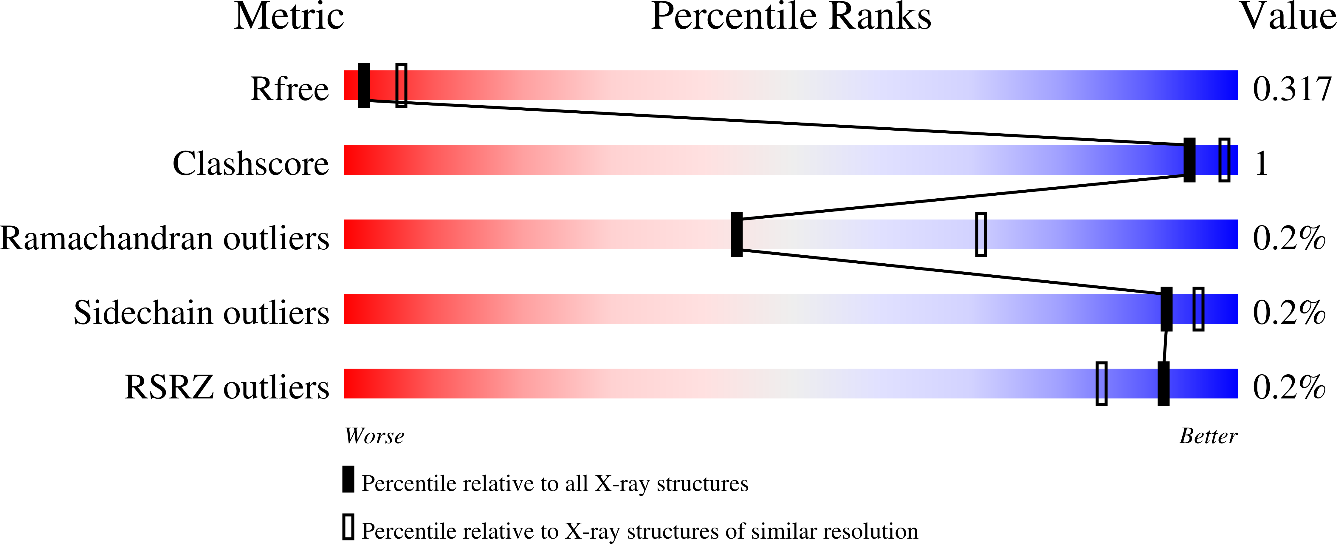
Resolution:
3.05 Å
R-Value Free:
0.31
R-Value Work:
0.26
R-Value Observed:
0.26
Space Group:
C 2 2 21
Macromolecular Entities
Structures with similar UniProt ID
Protein Blast
Polymer Type:polypeptide(L)
Molecule:Putative transposon Tn552 DNA
Gene (Uniprot):bin3
Mutagens:R54E, I100T
Chain IDs:A (auth: B), B (auth: D), C (auth: A), D (auth: C)
Chain Length:128
Number of Molecules:4
Biological Source:Staphylococcus aureus
Modified Residue
Compound ID Chain ID Parent Comp ID Details 2D Image
MSE A MET modified residue
Ligand Molecules
Primary Citation
Snapshots of a molecular swivel in action.
Nucleic Acids Res 46 5286 ? (2018)
PMID: 29315406 DOI: .

Abstact

Members of the serine family of site-specific recombinases exchange DNA strands via 180° rotation about a central protein-protein interface. Modeling of this process has been hampered by the lack of structures in more than one rotational state for any individual serine recombinase. Here we report crystal structures of the catalytic domains of four constitutively active mutants of the serine recombinase Sin, providing snapshots of rotational states not previously visualized for Sin, including two seen in the same crystal. Normal mode analysis predicted that each tetramer's lowest frequency mode (i.e. most accessible large-scale motion) mimics rotation: two protomers rotate as a pair with respect to the other two. Our analyses also suggest that rotation is not a rigid body movement around a single symmetry axis but instead uses multiple pivot points and entails internal motions within each subunit.

Legend

Protein

Chemical

Disease

Primary Citation of related structures
Feedback Form
Name
Email
Institute
Feedback