4WNK image
Deposition Date 2014-10-13
Release Date 2015-06-10
Last Version Date 2023-09-27
Entry Detail
PDB ID:
4WNK
Keywords:
Title:
Crystal Structure of Bovine G Protein Coupled-Receptor Kinase 5 in Complex with CCG215022
Biological Source:
Source Organism(s):
Bos taurus (Taxon ID: 9913)
Expression System(s):
Method Details:
Experimental Method:
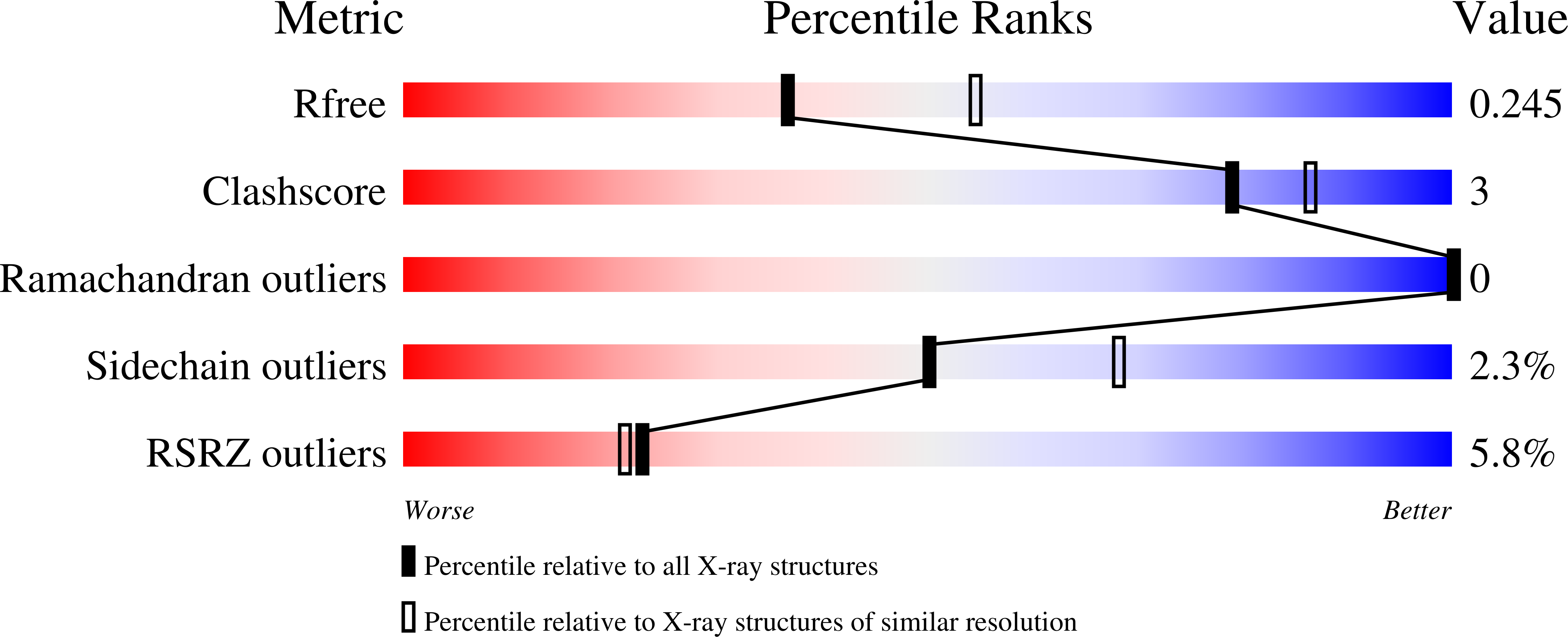
Resolution:
2.42 Å
R-Value Free:
0.24
R-Value Work:
0.19
R-Value Observed:
0.19
Space Group:
P 21 21 21
Macromolecular Entities
Structures with similar UniProt ID
Protein Blast
Polymer Type:polypeptide(L)
Molecule:G protein-coupled receptor ki
Gene (Uniprot):GRK5
Chain IDs:A
Chain Length:598
Number of Molecules:1
Biological Source:Bos taurus
Primary Citation
Crystal Structure of G Protein-coupled Receptor Kinase 5 in Complex with a Rationally Designed Inhibitor.
J. Biol. Chem. 290 20649 20659 (2015)
PMID: 26032411 DOI: 10.1074/jbc.M115.647370

Abstact

G protein-coupled receptor kinases (GRKs) regulate cell signaling by initiating the desensitization of active G protein-coupled receptors. The two most widely expressed GRKs (GRK2 and GRK5) play a role in cardiovascular disease and thus represent important targets for the development of novel therapeutic drugs. In the course of a GRK2 structure-based drug design campaign, one inhibitor (CCG215022) exhibited nanomolar IC50 values against both GRK2 and GRK5 and good selectivity against other closely related kinases such as GRK1 and PKA. Treatment of murine cardiomyocytes with CCG215022 resulted in significantly increased contractility at 20-fold lower concentrations than paroxetine, an inhibitor with more modest selectivity for GRK2. A 2.4 Å crystal structure of the GRK5·CCG215022 complex was determined and revealed that the inhibitor binds in the active site similarly to its parent compound GSK180736A. As designed, its 2-pyridylmethyl amide side chain occupies the hydrophobic subsite of the active site where it forms three additional hydrogen bonds, including one with the catalytic lysine. The overall conformation of the GRK5 kinase domain is similar to that of a previously determined structure of GRK6 in what is proposed to be its active state, but the C-terminal region of the enzyme adopts a distinct conformation. The kinetic properties of site-directed mutants in this region are consistent with the hypothesis that this novel C-terminal structure is representative of the membrane-bound conformation of the enzyme.

Legend

Protein

Chemical

Disease

Primary Citation of related structures
Feedback Form
Name
Email
Institute
Feedback