Abstact
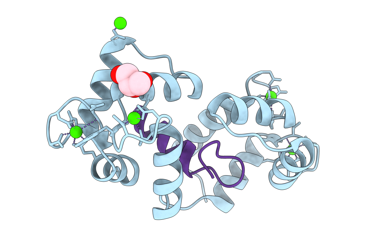
IP3-3K [Ins(1,4,5)P3 3-kinase] is a key enzyme that catalyses the synthesis of Ins(1,3,4,5)P4, using Ins(1,4,5)P3 and ATP as substrates. Both inositides, substrate and product, present crucial roles in the cell. Ins(1,4,5)P3 is a key point in Ca2+ metabolism that promotes Ca2+ release from intracellular stores and together with Ins(1,3,4,5)P4 regulates Ca2+ homoeostasis. In addition, Ins(1,3,4,5)P4 is involved in immune cell development. It has been proved that Ca2+/CaM (calmodulin) regulates the activity of IP3-3K, via direct interaction between both enzymes. Although we have extensive structural knowledge of the kinase domains of the three IP3-3K isoforms, no structural information is available about the interaction between IP3-3K and Ca2+/CaM. In the present paper we describe the crystal structure of the complex between human Ca2+/CaM and the CaM-binding region of human IP3-3K isoform A (residues 158-183) and propose a model for a complex including the kinase domain. The structure obtained allowed us to identify all of the key residues involved in the interaction, which have been evaluated by site-directed mutagenesis, pull-down and fluorescence anisotropy experiments. The results allowed the identification of a new CaM-binding motif, expanding our knowledge about how CaM interacts with its partners.