4IIT image
Deposition Date 2012-12-20
Release Date 2013-11-06
Last Version Date 2023-09-20
Entry Detail
PDB ID:
4IIT
Keywords:
Title:
The Phenylacetyl-CoA monooxygenase PaaABC subcomplex with phenylacetyl-CoA
Biological Source:
Source Organism(s):
Expression System(s):
Method Details:
Experimental Method:
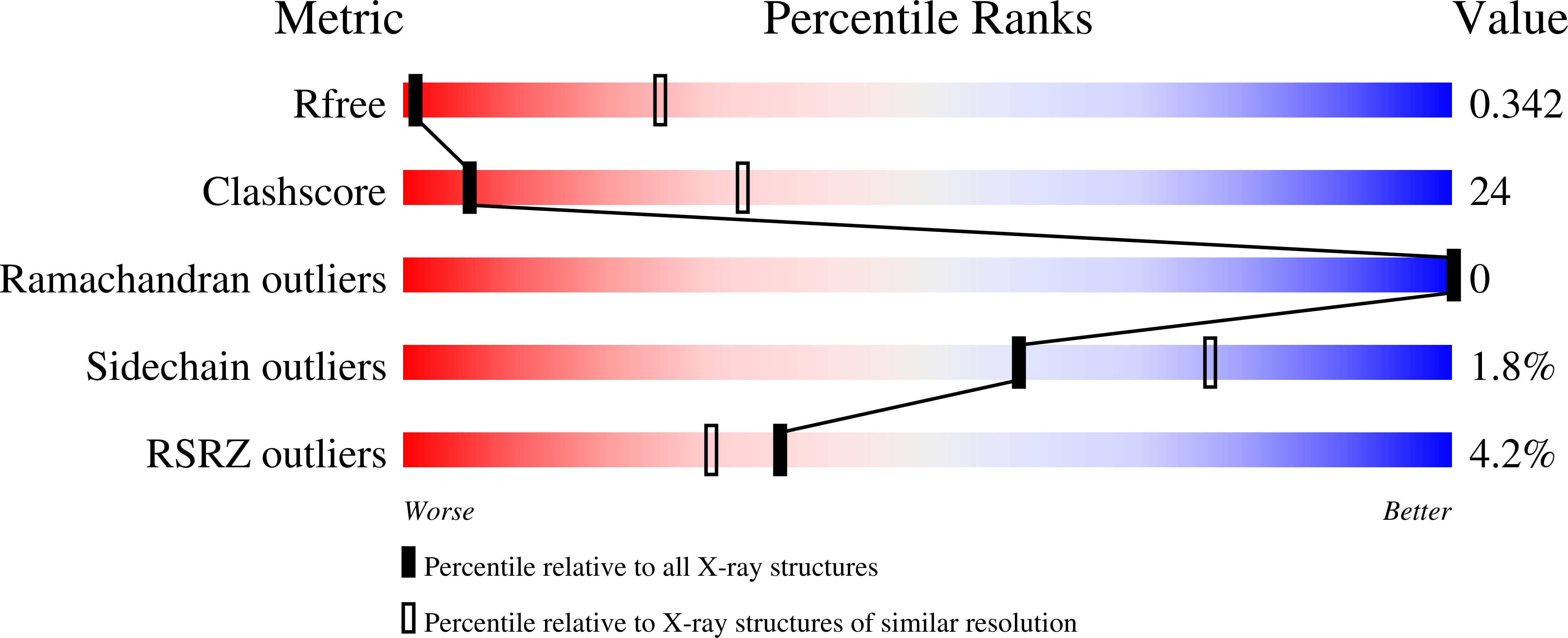
Resolution:
4.30 Å
R-Value Free:
0.36
R-Value Work:
0.28
R-Value Observed:
0.29
Space Group:
P 63 2 2
Macromolecular Entities
Structures with similar UniProt ID
Protein Blast
Polymer Type:polypeptide(L)
Molecule:Phenylacetate-CoA oxygenase s
Gene (Uniprot):paaA
Chain IDs:A
Chain Length:320
Number of Molecules:1
Biological Source:Klebsiella pneumoniae subsp. pneumoniae
Structures with similar UniProt ID
Protein Blast
Polymer Type:polypeptide(L)
Molecule:Phenylacetate-CoA oxygenase s
Gene (Uniprot):paaB
Chain IDs:B
Chain Length:95
Number of Molecules:1
Biological Source:Klebsiella pneumoniae subsp. pneumoniae
Structures with similar UniProt ID
Protein Blast
Polymer Type:polypeptide(L)
Molecule:Phenylacetate-CoA oxygenase s
Gene (Uniprot):paaC
Chain IDs:C
Chain Length:251
Number of Molecules:1
Biological Source:Klebsiella pneumoniae subsp. pneumoniae
Ligand Molecules
Primary Citation
Family of phenylacetyl-CoA monooxygenases differs in subunit organization from other monooxygenases.
J. Struct. Biol. 184 147 154 (2013)
PMID: 24055609 DOI: 10.1016/j.jsb.2013.09.012

Abstact

The phenylacetate degradation pathway is present in a wide range of microbes. A key component of this pathway is the four-subunit phenylacetyl-coenzyme A monooxygenase complex (PA-CoA MO, PaaACBE) that catalyzes the insertion of an oxygen in the aromatic ring of PA. This multicomponent enzyme represents a new family of monooxygenases. We have previously determined the structure of the PaaAC subcomplex of catalytic (A) and structural (C) subunits and shown that PaaACB form a stable complex. The PaaB subunit is unrelated to the small subunits of homologous monooxygenases and its role and organization of the PaaACB complex is unknown. From low-resolution crystal structure, electron microscopy and small angle X-ray scattering we show that the PaaACB complex forms heterohexamers, with a homodimer of PaaB bridging two PaaAC heterodimers. Modeling the interactions of reductase subunit PaaE with PaaACB suggested that a unique and conserved 'lysine bridge' constellation near the Fe-binding site in the PaaA subunit (Lys68, Glu49, Glu72 and Asp126) may form part of the electron transfer path from PaaE to the iron center. The crystal structure of the PaaA(K68Q/E49Q)-PaaC is very similar to the wild-type enzyme structure, but when combined with the PaaE subunit the mutant showed 20-50 times reduced activity, supporting the functional importance of the 'lysine bridge'.

Legend

Protein

Chemical

Disease

Primary Citation of related structures
Feedback Form
Name
Email
Institute
Feedback