4DHJ image
Deposition Date 2012-01-27
Release Date 2012-02-22
Last Version Date 2025-03-26
Entry Detail
PDB ID:
4DHJ
Title:
The structure of a ceOTUB1 ubiquitin aldehyde UBC13~Ub complex
Biological Source:
Source Organism(s):
Expression System(s):
Method Details:
Experimental Method:
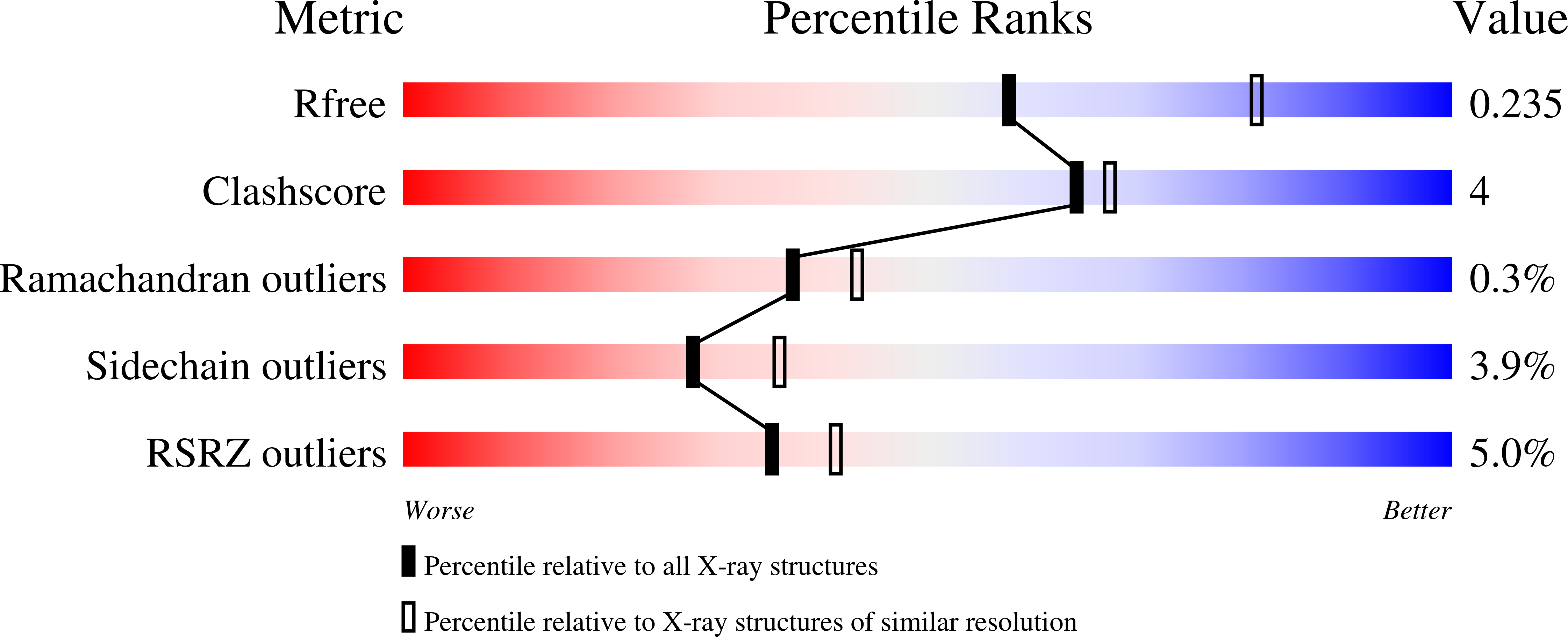
Resolution:
2.35 Å
R-Value Free:
0.23
R-Value Work:
0.20
R-Value Observed:
0.20
Space Group:
P 21 21 21
Macromolecular Entities
Structures with similar UniProt ID
Protein Blast
Polymer Type:polypeptide(L)
Molecule:Ubiquitin thioesterase otubai
Gene (Uniprot):otub-1
Chain IDs:A, C (auth: E), G (auth: I), K (auth: L)
Chain Length:284
Number of Molecules:4
Biological Source:Caenorhabditis elegans
Structures with similar UniProt ID
Protein Blast
Polymer Type:polypeptide(L)
Molecule:Ubiquitin aldehyde
Gene (Uniprot):UBC
Chain IDs:B, D (auth: F), H (auth: J), L (auth: M)
Chain Length:76
Number of Molecules:4
Biological Source:Homo sapiens
Structures with similar UniProt ID
Protein Blast
Polymer Type:polypeptide(L)
Molecule:Ubiquitin-conjugating enzyme
Gene (Uniprot):UBE2N
Chain IDs:F (auth: C), I (auth: K), J (auth: G), N
Chain Length:152
Number of Molecules:4
Biological Source:Homo sapiens
Structures with similar UniProt ID
Protein Blast
Polymer Type:polypeptide(L)
Molecule:Ubiquitin
Gene (Uniprot):UBC
Mutagens:G76C
Chain IDs:E (auth: D), M (auth: H)
Chain Length:76
Number of Molecules:2
Biological Source:Homo sapiens
Modified Residue
Compound ID Chain ID Parent Comp ID Details 2D Image
GLZ B GLY AMINO-ACETALDEHYDE
Primary Citation
The mechanism of OTUB1-mediated inhibition of ubiquitination.
Nature 483 618 622 (2012)
PMID: 22367539 DOI: 10.1038/nature10911

Abstact

Histones are ubiquitinated in response to DNA double-strand breaks (DSB), promoting recruitment of repair proteins to chromatin. UBC13 (also known as UBE2N) is a ubiquitin-conjugating enzyme (E2) that heterodimerizes with UEV1A (also known as UBE2V1) and synthesizes K63-linked polyubiquitin (K63Ub) chains at DSB sites in concert with the ubiquitin ligase (E3), RNF168 (ref. 3). K63Ub synthesis is regulated in a non-canonical manner by the deubiquitinating enzyme, OTUB1 (OTU domain-containing ubiquitin aldehyde-binding protein 1), which binds preferentially to the UBC13∼Ub thiolester. Residues amino-terminal to the OTU domain, which had been implicated in ubiquitin binding, are required for binding to UBC13∼Ub and inhibition of K63Ub synthesis. Here we describe structural and biochemical studies elucidating how OTUB1 inhibits UBC13 and other E2 enzymes. We unexpectedly find that OTUB1 binding to UBC13∼Ub is allosterically regulated by free ubiquitin, which binds to a second site in OTUB1 and increases its affinity for UBC13∼Ub, while at the same time disrupting interactions with UEV1A in a manner that depends on the OTUB1 N terminus. Crystal structures of an OTUB1-UBC13 complex and of OTUB1 bound to ubiquitin aldehyde and a chemical UBC13∼Ub conjugate show that binding of free ubiquitin to OTUB1 triggers conformational changes in the OTU domain and formation of a ubiquitin-binding helix in the N terminus, thus promoting binding of the conjugated donor ubiquitin in UBC13∼Ub to OTUB1. The donor ubiquitin thus cannot interact with the E2 enzyme, which has been shown to be important for ubiquitin transfer. The N-terminal helix of OTUB1 is positioned to interfere with UEV1A binding to UBC13, as well as with attack on the thiolester by an acceptor ubiquitin, thereby inhibiting K63Ub synthesis. OTUB1 binding also occludes the RING E3 binding site on UBC13, thus providing a further component of inhibition. The general features of the inhibition mechanism explain how OTUB1 inhibits other E2 enzymes in a non-catalytic manner.

Legend

Protein

Chemical

Disease

Primary Citation of related structures
Feedback Form
Name
Email
Institute
Feedback