Deposition Date
2012-04-27
Release Date
2012-08-22
Last Version Date
2024-11-20
Entry Detail
PDB ID:
4AS2
Keywords:
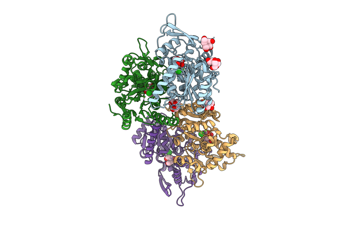
Title:
Pseudomonas Aeruginosa Phosphorylcholine Phosphatase. Monoclinic form
Biological Source:
Source Organism(s):
PSEUDOMONAS AERUGINOSA (Taxon ID: 287)
Expression System(s):
Method Details:
Experimental Method:
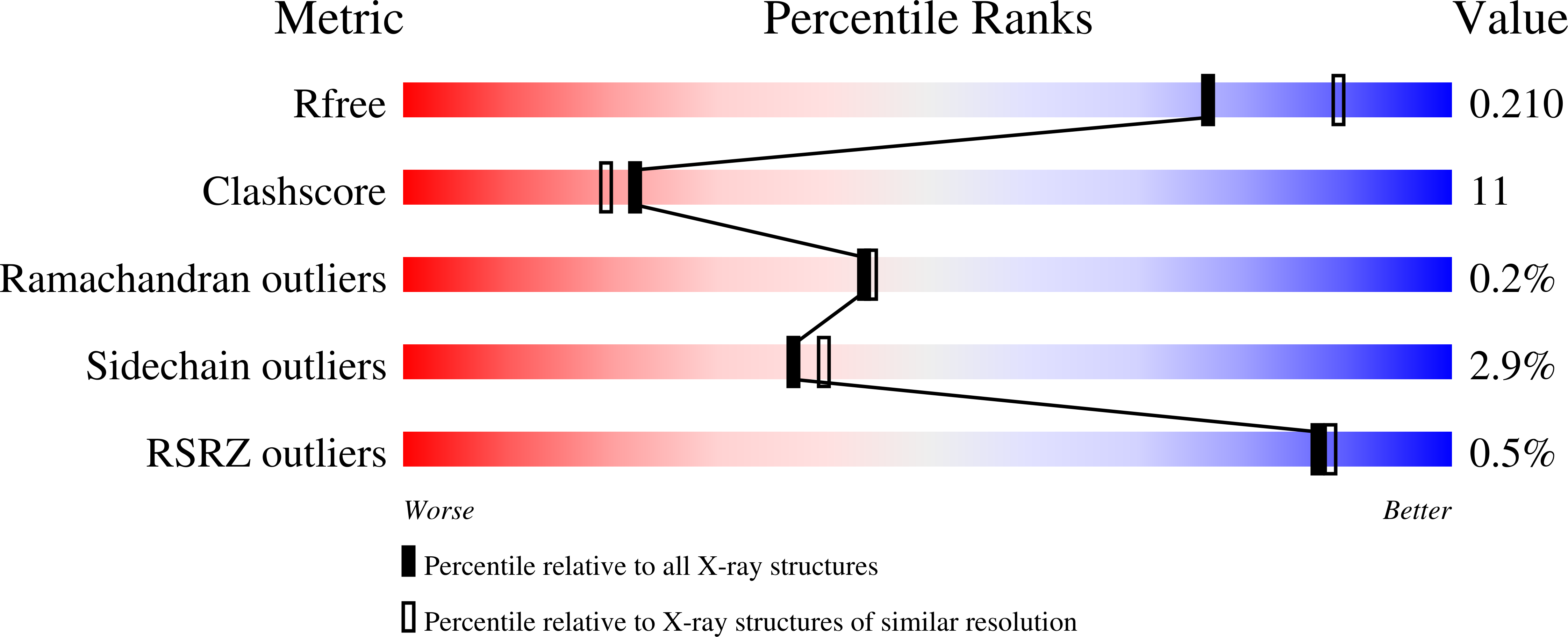
Resolution:
2.12 Å
R-Value Free:
0.21
R-Value Work:
0.15
R-Value Observed:
0.15
Space Group:
C 1 2 1