4AL9 image
Deposition Date 2012-03-02
Release Date 2013-03-20
Last Version Date 2026-01-21
Entry Detail
PDB ID:
4AL9
Title:
Crystal structure of the lectin PA-IL from Pseudomonas aeruginoas in complex with melibiose
Biological Source:
Source Organism(s):
Expression System(s):
Method Details:
Experimental Method:
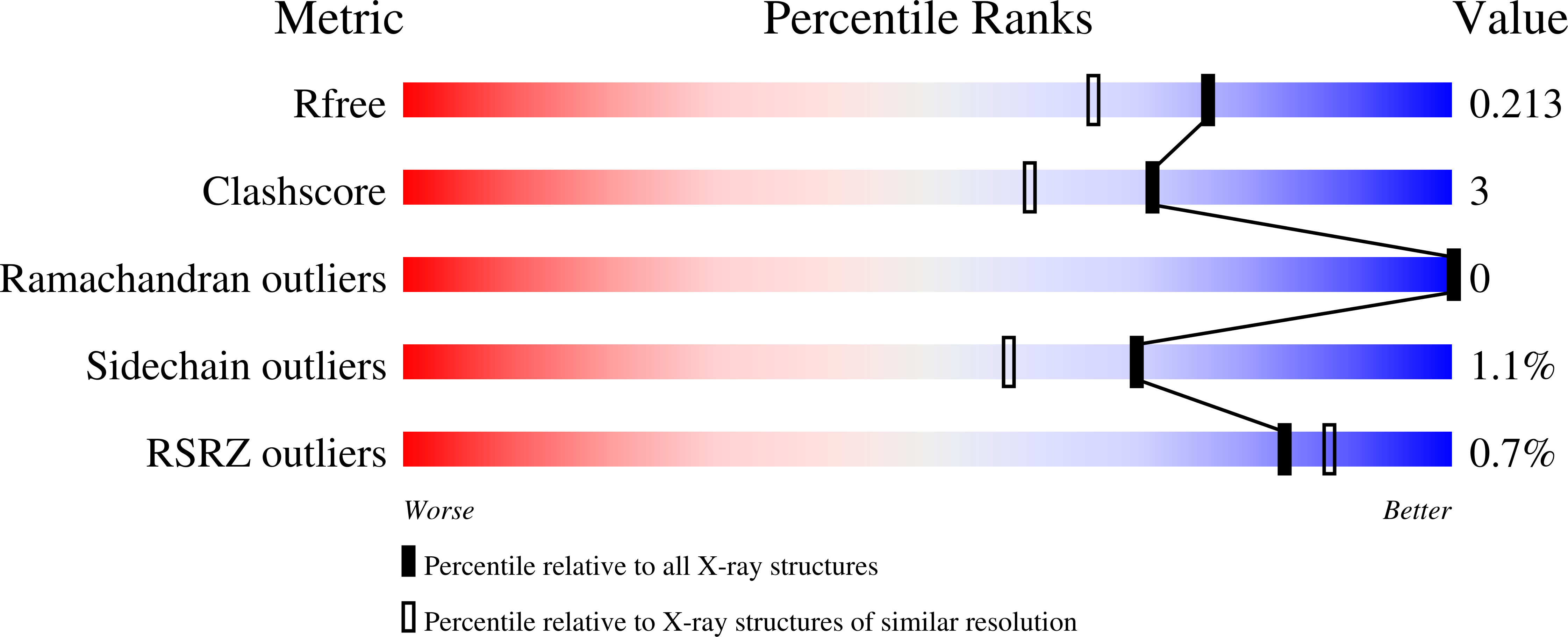
Resolution:
1.75 Å
R-Value Free:
0.21
R-Value Work:
0.16
R-Value Observed:
0.16
Space Group:
P 1
Macromolecular Entities
Structures with similar UniProt ID
Protein Blast
Polymer Type:polypeptide(L)
Molecule:PA-I GALACTOPHILIC LECTIN
Gene (Uniprot):lecA
Chain IDs:A, B, C, D, E, F, G, H
Chain Length:121
Number of Molecules:8
Biological Source:PSEUDOMONAS AERUGINOSA
Peptide-like Molecules
PRD_900037
Primary Citation
Secondary Sugar Binding Site Identified for Leca Lectin from Pseudomonas Aeruginosa.
Proteins 82 1060 ? (2014)
PMID: 24123124 DOI: 10.1002/PROT.24430

Abstact

The galactose-specific lectin LecA from Pseudomonas aeruginosa is a target for the development of new anti-infectious compounds. Sugar based molecules with anti-adhesive properties present great potential in the fight against bacterial infection and biofilm formation. LecA is specific for oligosaccharides with terminal α-galactoside residues and displays strong affinity for melibiose (αGal1-6Glc) with a Kd of 38.8 µM. The crystal structure of LecA/melibiose complex shows classical calcium-bridged binding of αGal in the primary binding site but also revealed a secondary sugar binding site with glucose bound. This sugar binding site is in close proximity to the galactose binding one, is independent of calcium and mainly involves interactions with a symmetry-related protein. This discovery would help to the design of new potent inhibitors targeting both binding sites.

Legend

Protein

Chemical

Disease

Primary Citation of related structures
Feedback Form
Name
Email
Institute
Feedback