3ZMI image
Deposition Date 2013-02-11
Release Date 2013-05-22
Last Version Date 2024-11-06
Entry Detail
PDB ID:
3ZMI
Keywords:
Title:
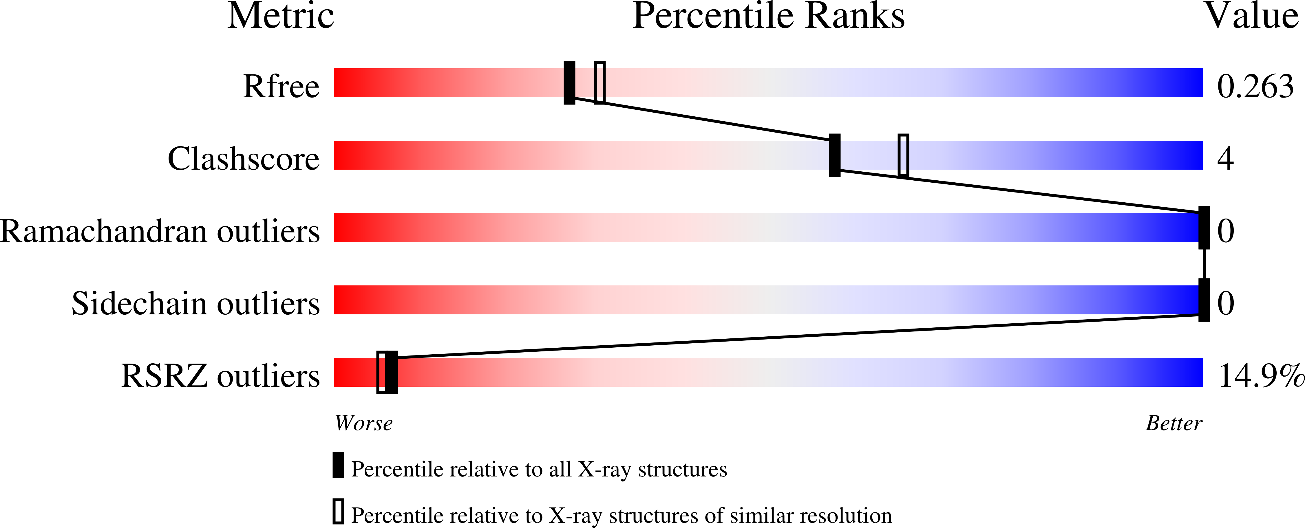
Structure of E.coli rhomboid protease GlpG in complex with monobactam L29
Biological Source:
Source Organism(s):
ESCHERICHIA COLI (Taxon ID: 562)
Expression System(s):
Method Details:
Experimental Method:
Resolution:
2.20 Å
R-Value Free:
0.26
R-Value Work:
0.21
R-Value Observed:
0.21
Space Group:
H 3 2
Macromolecular Entities
Structures with similar UniProt ID
Protein Blast
Polymer Type:polypeptide(L)
Molecule:RHOMBOID PROTEASE GLPG
Gene (Uniprot):glpG
Chain IDs:A
Chain Length:179
Number of Molecules:1
Biological Source:ESCHERICHIA COLI
Primary Citation
Structure of Rhomboid Protease in Complex with Beta-Lactam Inhibitors Defines the S2' Cavity.
Structure 21 1051 1058 (2013)
PMID: 23665170 DOI: 10.1016/J.STR.2013.03.013

Abstact

Rhomboids are evolutionarily conserved serine proteases that cleave transmembrane proteins within the membrane. The increasing number of known rhomboid functions in prokaryotes and eukaryotes makes them attractive drug targets. Here, we describe structures of the Escherichia coli rhomboid GlpG in complex with β-lactam inhibitors. The inhibitors form a single bond to the catalytic serine and the carbonyl oxygen of the inhibitor faces away from the oxyanion hole. The hydrophobic N-substituent of β-lactam inhibitors points into a cavity within the enzyme, providing a structural explanation for the specificity of β-lactams on rhomboid proteases. This same cavity probably represents the S2' substrate binding site of GlpG. We suggest that the structural changes in β-lactam inhibitor binding reflect the state of the enzyme at an initial stage of substrate binding to the active site. The structural insights from these enzyme-inhibitor complexes provide a starting point for structure-based design for rhomboid inhibitors.

Legend

Protein

Chemical

Disease

Primary Citation of related structures
Feedback Form
Name
Email
Institute
Feedback