

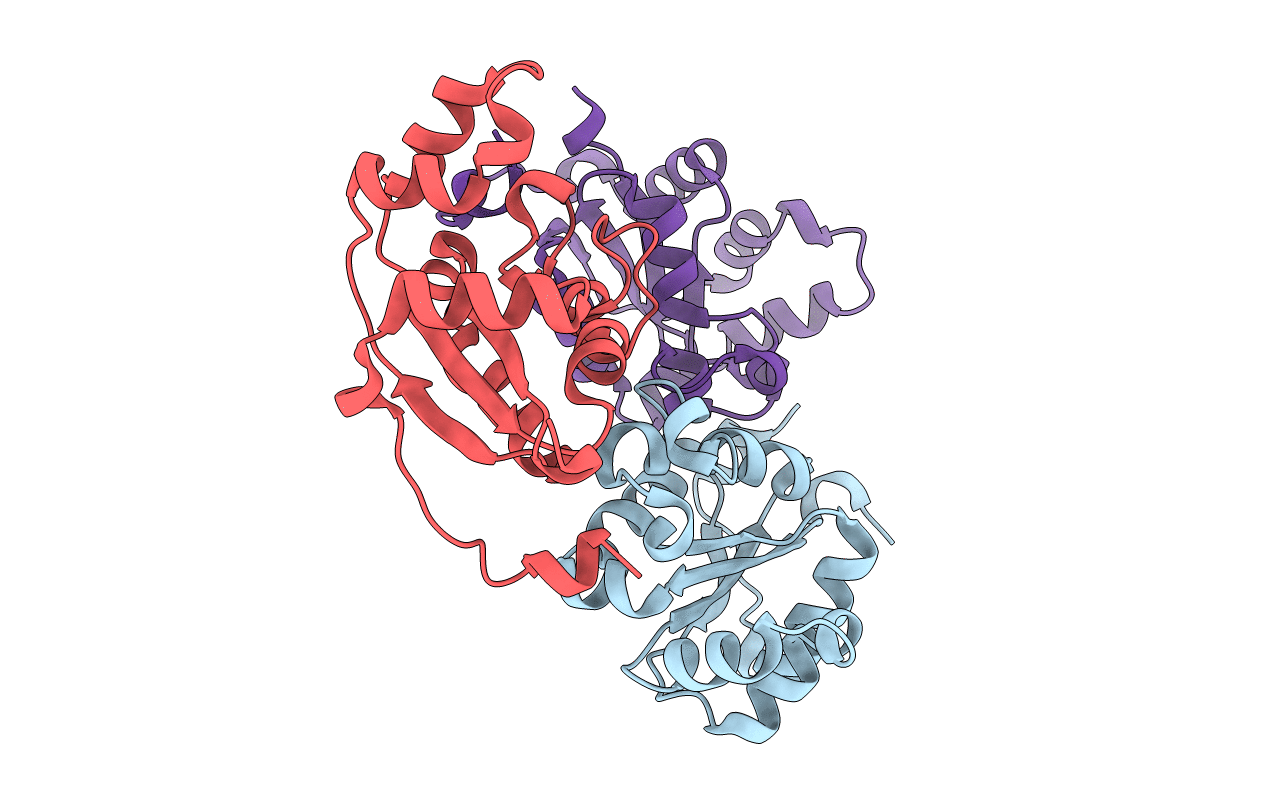
3WX8 image
Deposition Date 2014-07-25
Release Date 2015-06-10
Last Version Date 2024-03-20
Entry Detail
PDB ID:
3WX8
Keywords:
Title:
Purification, characterization and structure of nucleoside diphosphate kinase from Drosophila S2 cells
Biological Source:
Source Organism(s):
Expression System(s):
Method Details:
Experimental Method:
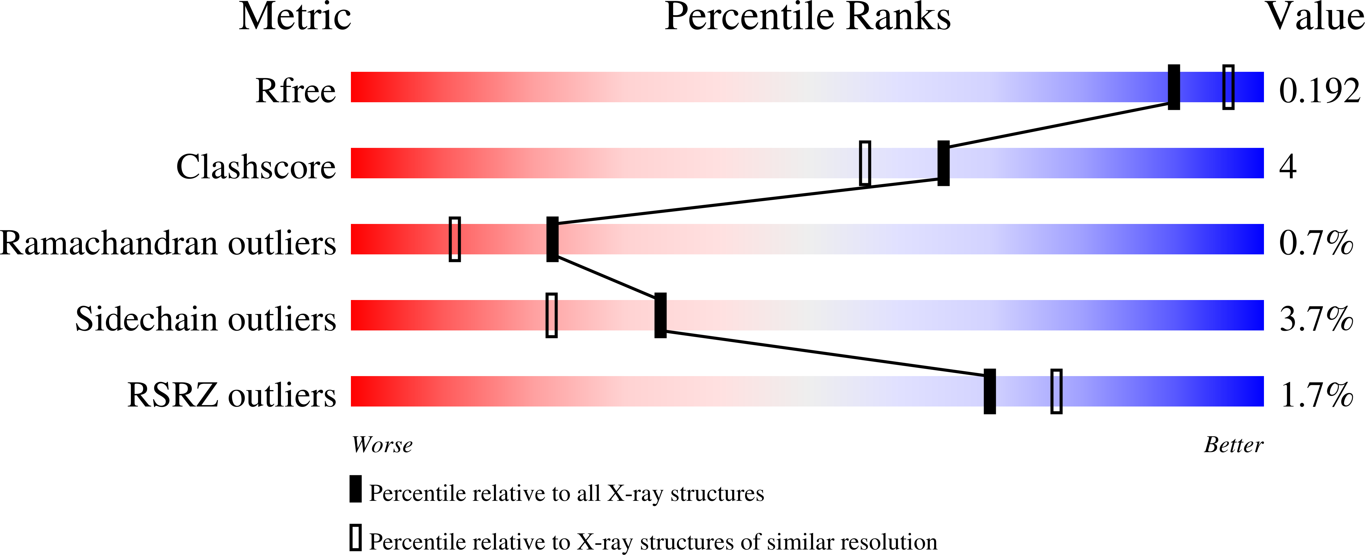
Resolution:
1.95 Å
R-Value Free:
0.18
R-Value Work:
0.15
R-Value Observed:
0.15
Space Group:
P 32 2 1
Macromolecular Entities
Structures with similar UniProt ID
Protein Blast
Polymer Type:polypeptide(L)
Molecule:Nucleoside diphosphate kinase
Gene (Uniprot):awd
Chain IDs:A, B, C
Chain Length:153
Number of Molecules:3
Biological Source:Drosophila melanogaster
Primary Citation
Purification, characterization and structure of nucleoside diphosphate kinase from Drosophila melanogaster
Protein Expr. Purif. 103 48 55 (2014)
PMID: 25195176 DOI: 10.1016/j.pep.2014.08.014

Abstact

Nucleoside diphosphate kinase (NDPK) is a ubiquitous enzyme found in all organisms and cell types, which catalyzes the transfer of the phosphoryl group from a nucleoside triphosphate to a nucleoside diphosphate. The gene encoding for NDPK from Drosophila melanogaster was amplified from the genomic DNA. The recombinant NDPK (rNDPK) was overexpressed in Escherichia coli and purified to homogeneity by Ni-NTA agarose affinity chromatography, HiTrap SP HP cation exchange chromatography and HiLoad 16/60 Superdex 200 gel filtration chromatography. The gel filtration chromatography and analytical ultracentrifugation showed that rNDPK was a trimer in solution. The binding affinity of NDPs with rNDPK, measured by isothermal titration calorimetry, indicated that the purines nucleotides show higher binding affinity compared with pyrimidines. The rNDPK had a definite nuclease activity in vitro, which could cleave supercoiled plasmid DNA, but had no effect on dsDNA and ssDNA. Furthermore, the structure for NDPK was determined by using the sitting drop vapor diffusion method. In the final model, the asymmetric unit is made of three molecules, each of which consists of a four-stranded anti-parallel β-sheets and seven α-helices. Sequence alignment and structure comparison illustrated that the simulated nucleotide-binding active site are conserved.

Legend

Protein

Chemical

Disease

Primary Citation of related structures
Feedback Form
Name
Email
Institute
Feedback