Deposition Date
2011-08-18
Release Date
2012-07-04
Last Version Date
2024-10-30
Entry Detail
PDB ID:
3VGP
Keywords:
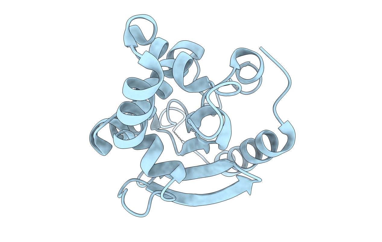
Title:
Crystal structure of the C-terminal globular domain of oligosaccharyltransferase (AF_0329) from Archaeoglobus fulgidus
Biological Source:
Source Organism(s):
Archaeoglobus fulgidus (Taxon ID: 224325)
Expression System(s):
Method Details:
Experimental Method:
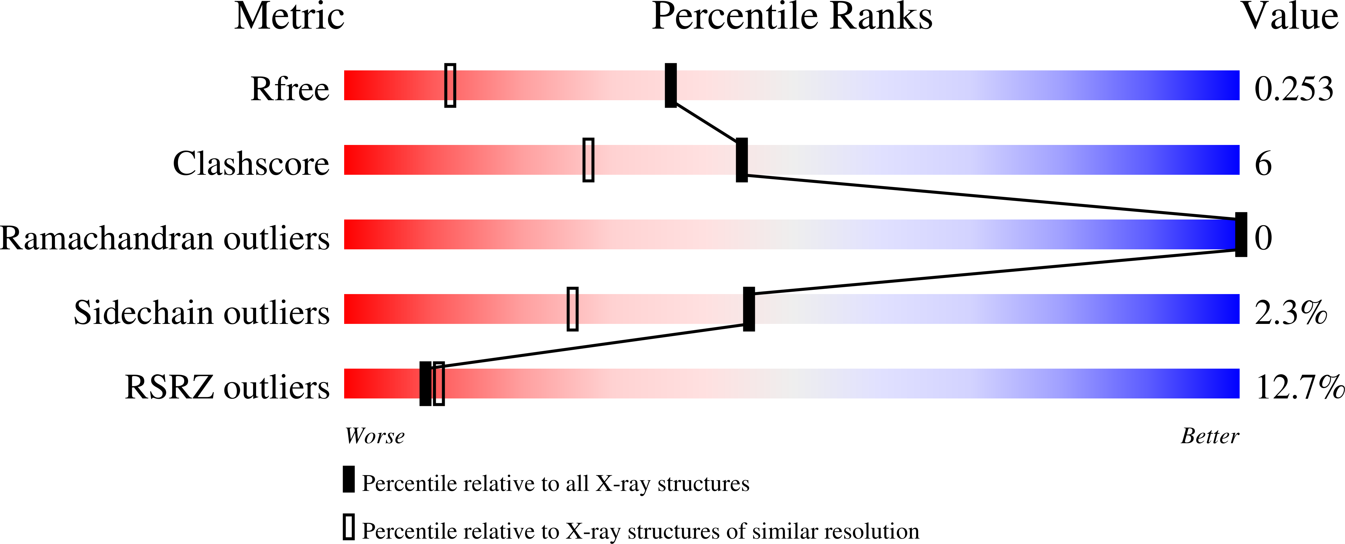
Resolution:
1.75 Å
R-Value Free:
0.25
R-Value Work:
0.22
R-Value Observed:
0.22
Space Group:
P 41 21 2