

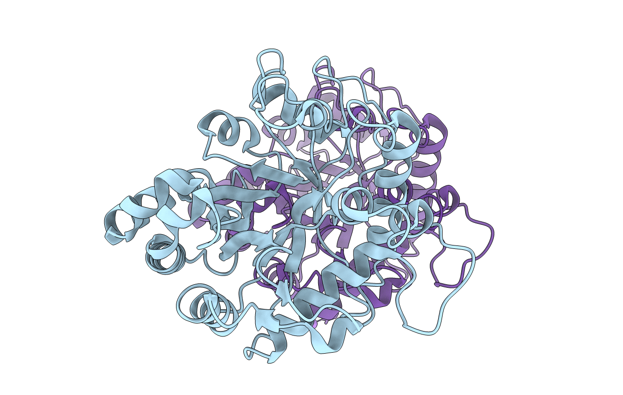
3UR7 image
Deposition Date 2011-11-21
Release Date 2012-05-30
Last Version Date 2023-09-13
Entry Detail
PDB ID:
3UR7
Keywords:
Title:
Higher-density crystal structure of potato endo-1,3-beta-glucanase
Biological Source:
Source Organism(s):
Expression System(s):
Method Details:
Experimental Method:
Resolution:
1.40 Å
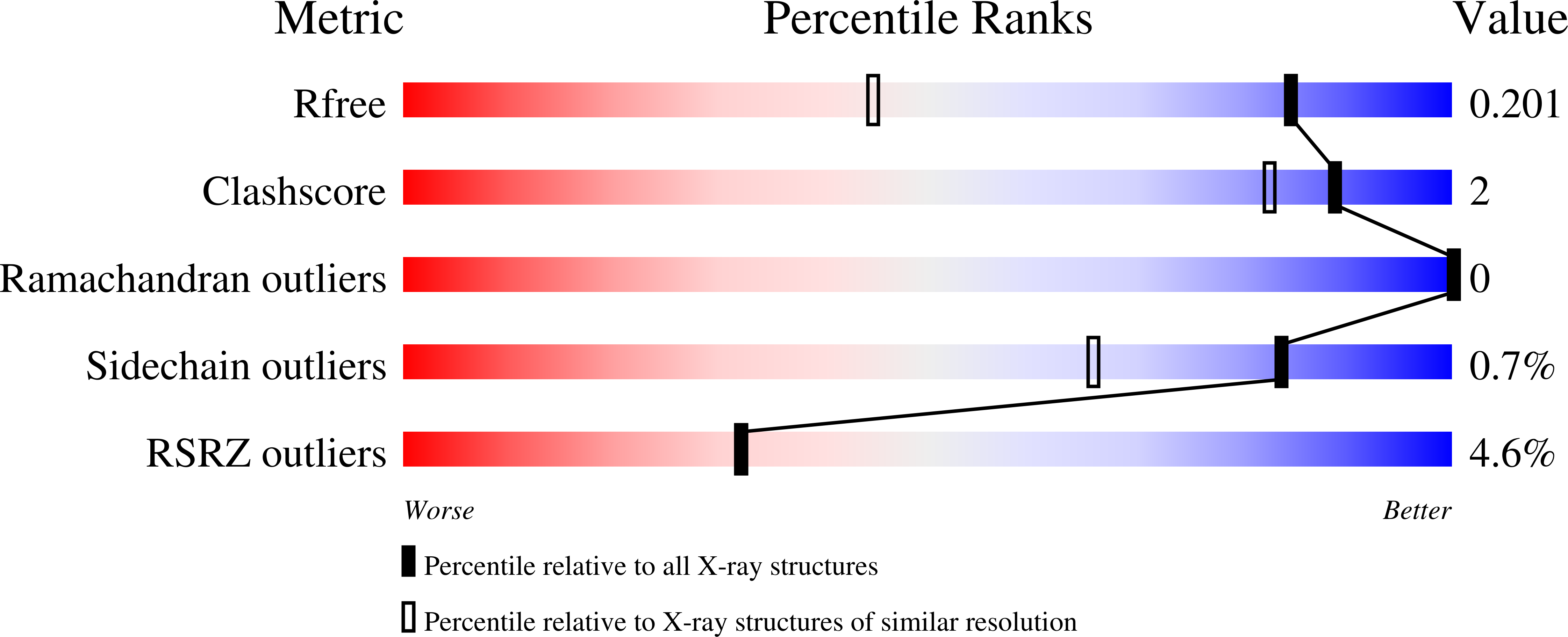
R-Value Free:
0.18
R-Value Work:
0.16
R-Value Observed:
0.16
Space Group:
P 1 21 1
Macromolecular Entities
Structures with similar UniProt ID
Protein Blast
Polymer Type:polypeptide(L)
Molecule:Glucan endo-1,3-beta-D-glucos
Gene (Uniprot):gluB20-2
Chain IDs:A, B
Chain Length:323
Number of Molecules:2
Biological Source:Solanum tuberosum
Ligand Molecules
Primary Citation
Two high-resolution structures of potato endo-1,3-beta-glucanase reveal subdomain flexibility with implications for substrate binding
Acta Crystallogr. D Biol. Crystallogr. 68 713 723 (2012)
PMID: 22683794 DOI: 10.1107/S090744491200995X

Abstact

Endo-1,3-β-glucanases are widely distributed among bacteria, fungi and higher plants. They are responsible for hydrolysis of the glycosidic bond in specific polysaccharides with tracts of unsubstituted β-1,3-linked glucosyl residues. The plant enzymes belong to glycoside hydrolase family 17 (GH17) and are also members of class 2 of pathogenesis-related (PR) proteins. X-ray diffraction data were collected to 1.40 and 1.26 Å resolution from two crystals of endo-1,3-β-glucanase from Solanum tuberosum (potato, cultivar Désirée) which, despite having a similar packing framework, represented two separate crystal forms. In particular, they differed in the Matthews coefficient and are consequently referred to as higher density (HD; 1.40 Å resolution) and lower density (LD; 1.26 Å resolution) forms. The general fold of the protein resembles that of other known plant endo-1,3-β-glucanases and is defined by a (β/α)(8)-barrel with an additional subdomain built around the C-terminal half of the barrel. The structures revealed high flexibility of the subdomain, which forms part of the catalytic cleft. Comparison with structures of other GH17 endo-1,3-β-glucanases revealed differences in the arrangement of the secondary-structure elements in this region, which can be correlated with sequence variability and may suggest distinct substrate-binding patterns. The crystal structures revealed an unusual packing mode, clearly visible in the LD structure, caused by the presence of the C-terminal His(6) tag, which extends from the compact fold of the enzyme molecule and docks in the catalytic cleft of a neighbouring molecule. In this way, an infinite chain of His-tag-linked protein molecules is formed along the c direction.

Legend

Protein

Chemical

Disease

Primary Citation of related structures
Feedback Form
Name
Email
Institute
Feedback