

3SEO image
Deposition Date 2011-06-10
Release Date 2011-08-31
Last Version Date 2024-02-28
Entry Detail
PDB ID:
3SEO
Title:
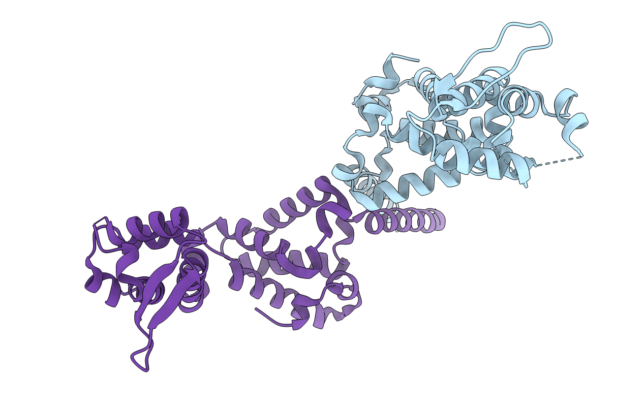
Crystal structure of VopL C terminal domain
Biological Source:
Source Organism(s):
Expression System(s):
Method Details:
Experimental Method:
Resolution:
2.31 Å
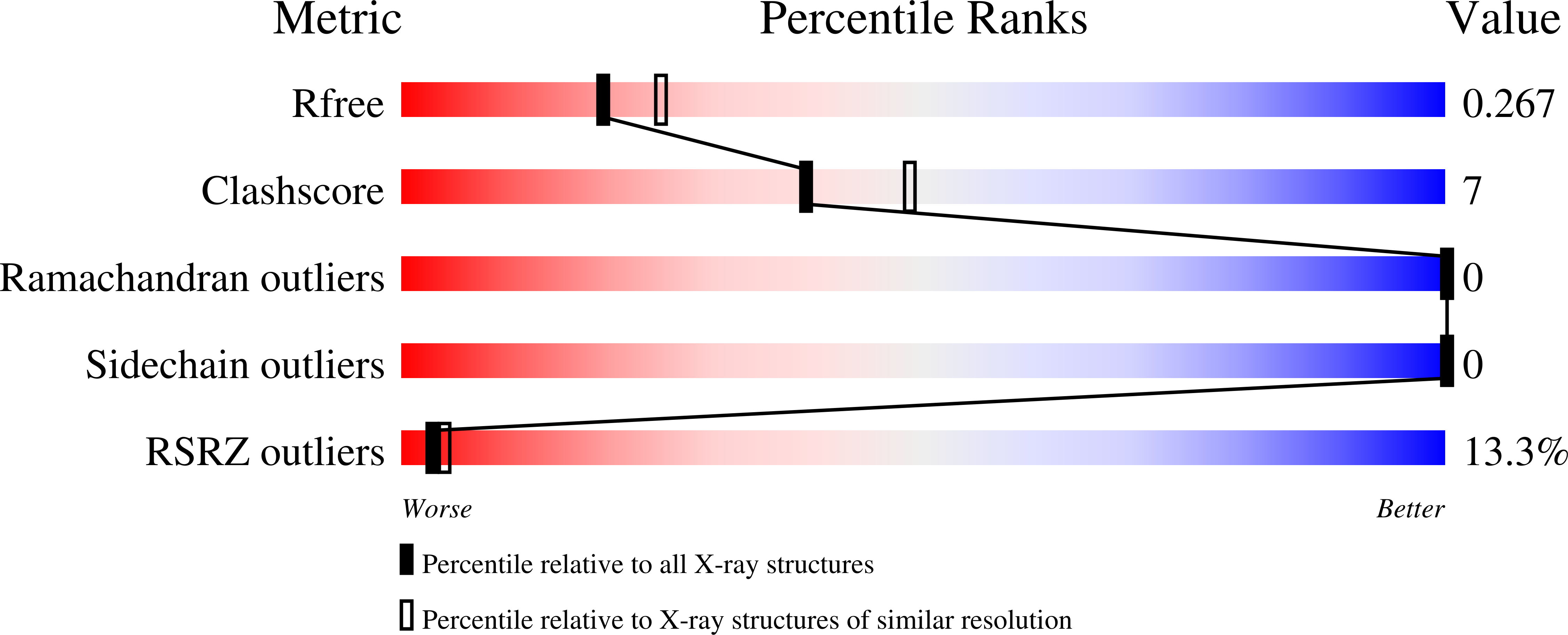
R-Value Free:
0.27
R-Value Work:
0.22
R-Value Observed:
0.22
Space Group:
P 21 21 21
Macromolecular Entities
Structures with similar UniProt ID
Protein Blast
Polymer Type:polypeptide(L)
Molecule:VopL C terminal domain protei
Gene (Uniprot):VPA1370
Chain IDs:A, B
Chain Length:241
Number of Molecules:2
Biological Source:Vibrio parahaemolyticus
Ligand Molecules
Primary Citation
Mechanism of actin filament nucleation by the bacterial effector VopL.
Nat. Struct. Mol. Biol. 18 1068 1074 (2011)
PMID: 21873984 DOI: 10.1038/nsmb.2110

Abstact

Vibrio parahaemolyticus protein L (VopL) is an actin nucleation factor that induces stress fibers when injected into eukaryotic host cells. VopL contains three N-terminal Wiskott-Aldrich homology 2 (WH2) motifs and a unique VopL C-terminal domain (VCD). We describe crystallographic and biochemical analyses of filament nucleation by VopL. The WH2 element of VopL does not nucleate on its own and requires the VCD for activity. The VCD forms a U-shaped dimer in the crystal, stabilized by a terminal coiled coil. Dimerization of the WH2 motifs contributes strongly to nucleation activity, as do contacts of the VCD to actin. Our data lead to a model in which VopL stabilizes primarily lateral (short-pitch) contacts between actin monomers to create the base of a two-stranded filament. Stabilization of lateral contacts may be a common feature of actin filament nucleation by WH2-based factors.

Legend

Protein

Chemical

Disease

Primary Citation of related structures
Feedback Form
Name
Email
Institute
Feedback