3S0Q image
Deposition Date 2011-05-13
Release Date 2011-08-31
Last Version Date 2024-02-28
Entry Detail
PDB ID:
3S0Q
Keywords:
Title:
Peptidase module of the peptidoglycan hydrolase RipA (Rv1477) from Mycobacterium tuberculosis, catalytic site mutant (Cys383Ala) at 1.45 resolution
Biological Source:
Source Organism(s):
Expression System(s):
Method Details:
Experimental Method:
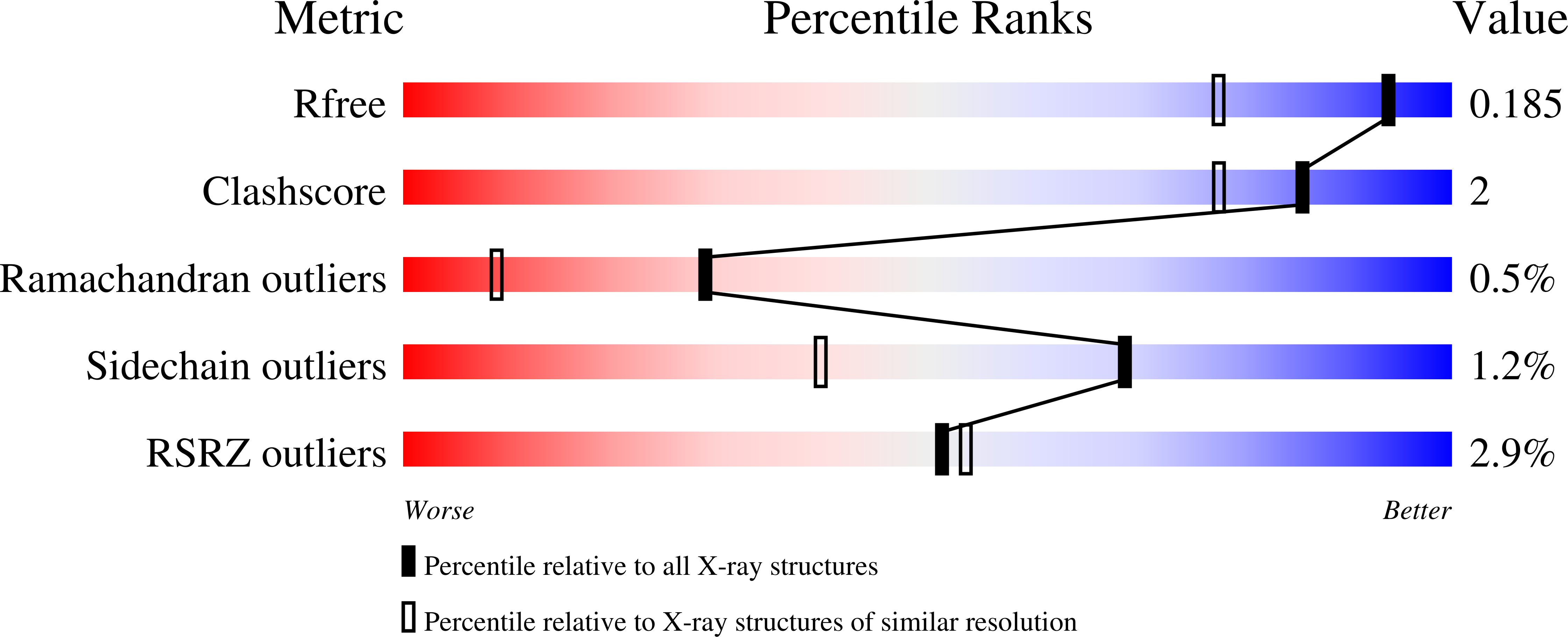
Resolution:
1.45 Å
R-Value Free:
0.17
R-Value Work:
0.15
R-Value Observed:
0.15
Space Group:
P 21 21 21
Macromolecular Entities
Structures with similar UniProt ID
Protein Blast
Polymer Type:polypeptide(L)
Molecule:INVASION PROTEIN
Gene (Uniprot):ripA
Mutagens:C383A
Chain IDs:A
Chain Length:215
Number of Molecules:1
Biological Source:Mycobacterium tuberculosis
Primary Citation
Peptidoglycan Remodeling in Mycobacterium tuberculosis: Comparison of Structures and Catalytic Activities of RipA and RipB.
J. Mol. Biol. 413 247 260 (2011)
PMID: 21864539 DOI: 10.1016/j.jmb.2011.08.014

Abstact

The success of Mycobacterium tuberculosis in sustaining long-term survival within the host macrophages partly relies on its unique cell envelop that also confers low susceptibility to several antibiotics. Remodeling of the septal peptidoglycan (PG) has been linked to the putative PG hydrolases RipA and RipB. The crystal structures of RipB (Rv1478) and the homologous module of RipA (Rv1477) were determined to 1.60 Å and 1.38 Å resolution, respectively. Both proteins contain a C-terminal core domain resembling the NlpC-type PG hydrolases. However, the structure of RipB exhibits striking differences to the structures of this domain in RipA reported here and previously by others. Major structural differences were found in the N-terminal segments of 70 amino acids and in an adjacent loop, which form part of the substrate binding groove. Both RipA and RipB are able to bind PG. RipA, its C-terminal module and RipB cleave defined PG fragments between d-glutamate and meso-diaminopimelate with pH optima of 5 and 6, respectively. The peptidase module of RipA is also able to degrade Bacillus subtilis PG, which displays peptide stems and cross-links identical with those found in mycobacterial murein. RipB did not show comparable hydrolase activity with this substrate. Removal of the N-terminal segments previously suggested to have a role in auto-inhibition did not change the activity of either RipA or RipB. A comparison of the putative active-site clefts in the two enzymes provides structural insights into the basis of the differences in their substrate specificity.

Legend

Protein

Chemical

Disease

Primary Citation of related structures
Feedback Form
Name
Email
Institute
Feedback