3RKO image
Deposition Date 2011-04-18
Release Date 2011-08-03
Last Version Date 2024-02-28
Entry Detail
PDB ID:
3RKO
Keywords:
Title:
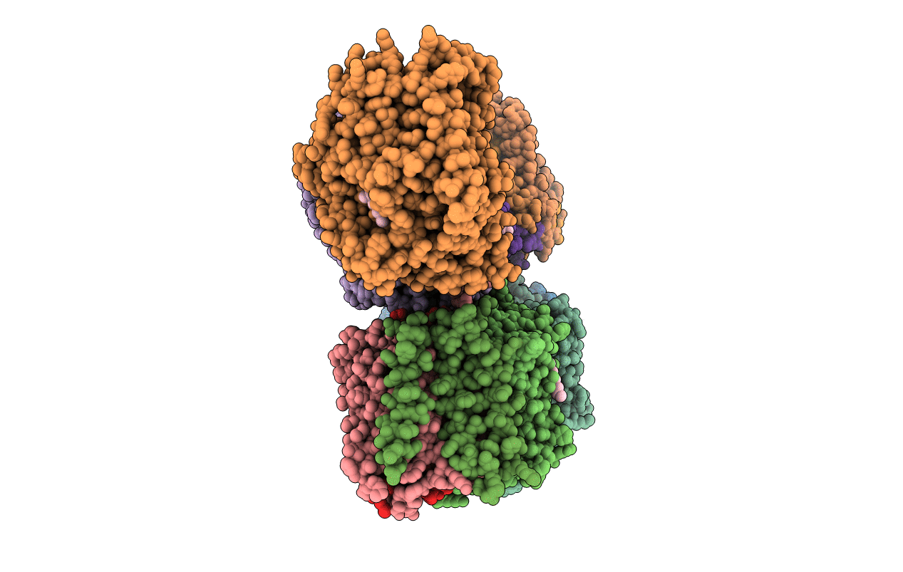
Crystal structure of the membrane domain of respiratory complex I from E. coli at 3.0 angstrom resolution
Biological Source:
Source Organism(s):
Method Details:
Experimental Method:
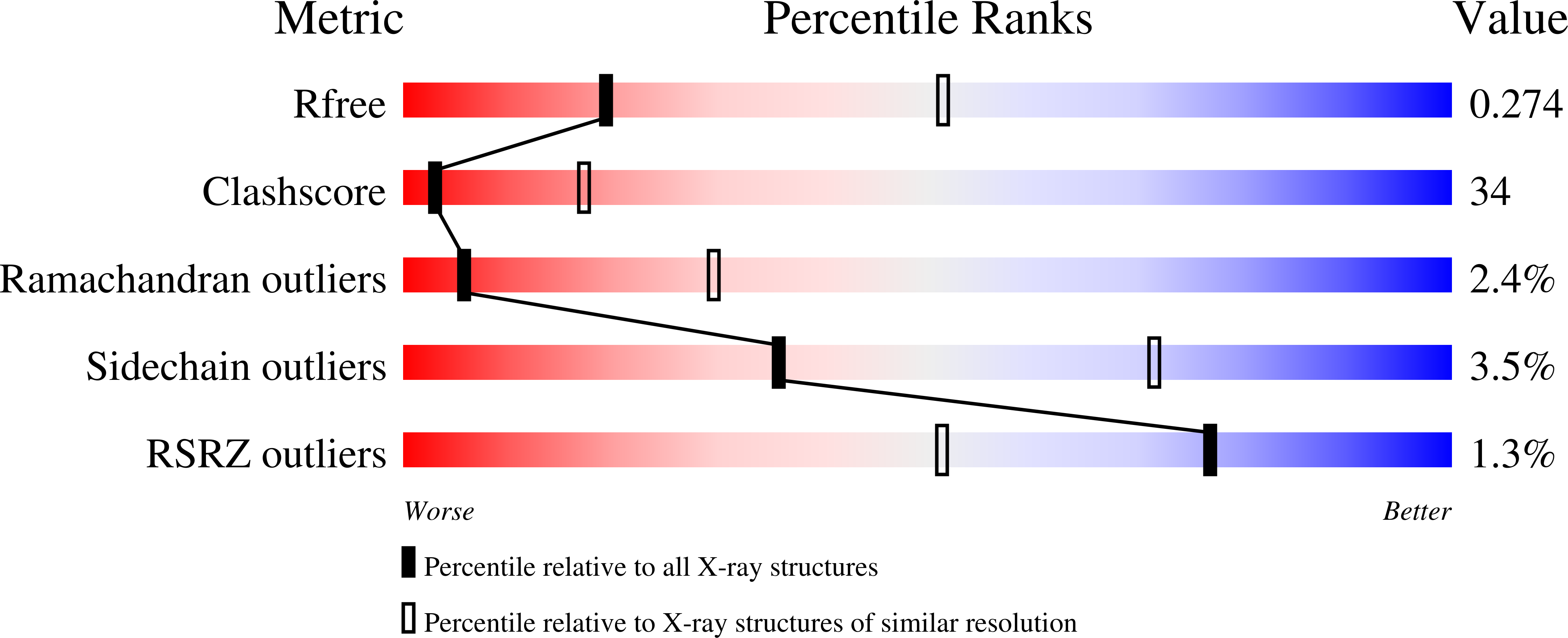
Resolution:
3.00 Å
R-Value Free:
0.28
R-Value Work:
0.23
R-Value Observed:
0.23
Space Group:
P 1
Macromolecular Entities
Structures with similar UniProt ID
Protein Blast
Polymer Type:polypeptide(L)
Molecule:NADH-QUINONE OXIDOREDUCTASE S
Chain IDs:E (auth: A), K (auth: E)
Chain Length:147
Number of Molecules:2
Biological Source:Escherichia coli
Structures with similar UniProt ID
Protein Blast
Polymer Type:polypeptide(L)
Molecule:NADH-QUINONE OXIDOREDUCTASE S
Chain IDs:F (auth: J), L (auth: F)
Chain Length:184
Number of Molecules:2
Biological Source:Escherichia coli
Structures with similar UniProt ID
Protein Blast
Polymer Type:polypeptide(L)
Molecule:NADH-QUINONE OXIDOREDUCTASE S
Chain IDs:D (auth: K), J (auth: G)
Chain Length:100
Number of Molecules:2
Biological Source:Escherichia coli
Structures with similar UniProt ID
Protein Blast
Polymer Type:polypeptide(L)
Molecule:NADH-QUINONE OXIDOREDUCTASE S
Chain IDs:A (auth: L), G (auth: B)
Chain Length:613
Number of Molecules:2
Biological Source:Escherichia coli
Structures with similar UniProt ID
Protein Blast
Polymer Type:polypeptide(L)
Molecule:NADH-QUINONE OXIDOREDUCTASE S
Chain IDs:B (auth: M), H (auth: C)
Chain Length:509
Number of Molecules:2
Biological Source:Escherichia coli
Structures with similar UniProt ID
Protein Blast
Polymer Type:polypeptide(L)
Molecule:NADH-QUINONE OXIDOREDUCTASE S
Chain IDs:C (auth: N), I (auth: D)
Chain Length:485
Number of Molecules:2
Biological Source:Escherichia coli
Primary Citation
Structure of the membrane domain of respiratory complex I.
Nature 476 414 420 (2011)
PMID: 21822288 DOI: 10.1038/nature10330

Abstact

Complex I is the first and largest enzyme of the respiratory chain, coupling electron transfer between NADH and ubiquinone to the translocation of four protons across the membrane. It has a central role in cellular energy production and has been implicated in many human neurodegenerative diseases. The L-shaped enzyme consists of hydrophilic and membrane domains. Previously, we determined the structure of the hydrophilic domain. Here we report the crystal structure of the Esherichia coli complex I membrane domain at 3.0 Å resolution. It includes six subunits, NuoL, NuoM, NuoN, NuoA, NuoJ and NuoK, with 55 transmembrane helices. The fold of the homologous antiporter-like subunits L, M and N is novel, with two inverted structural repeats of five transmembrane helices arranged, unusually, face-to-back. Each repeat includes a discontinuous transmembrane helix and forms half of a channel across the membrane. A network of conserved polar residues connects the two half-channels, completing the proton translocation pathway. Unexpectedly, lysines rather than carboxylate residues act as the main elements of the proton pump in these subunits. The fourth probable proton-translocation channel is at the interface of subunits N, K, J and A. The structure indicates that proton translocation in complex I, uniquely, involves coordinated conformational changes in six symmetrical structural elements.

Legend

Protein

Chemical

Disease

Primary Citation of related structures
Feedback Form
Name
Email
Institute
Feedback