3RJF image
Deposition Date 2011-04-15
Release Date 2012-01-18
Last Version Date 2023-09-13
Entry Detail
PDB ID:
3RJF
Title:
Ternary complex of DNA Polymerase Beta with a gapped DNA containing (syn)8odG at template position paired with non-hydrolyzable dATP analog (dApCPP)
Biological Source:
Source Organism(s):
Homo sapiens (Taxon ID: 9606)
Expression System(s):
Method Details:
Experimental Method:
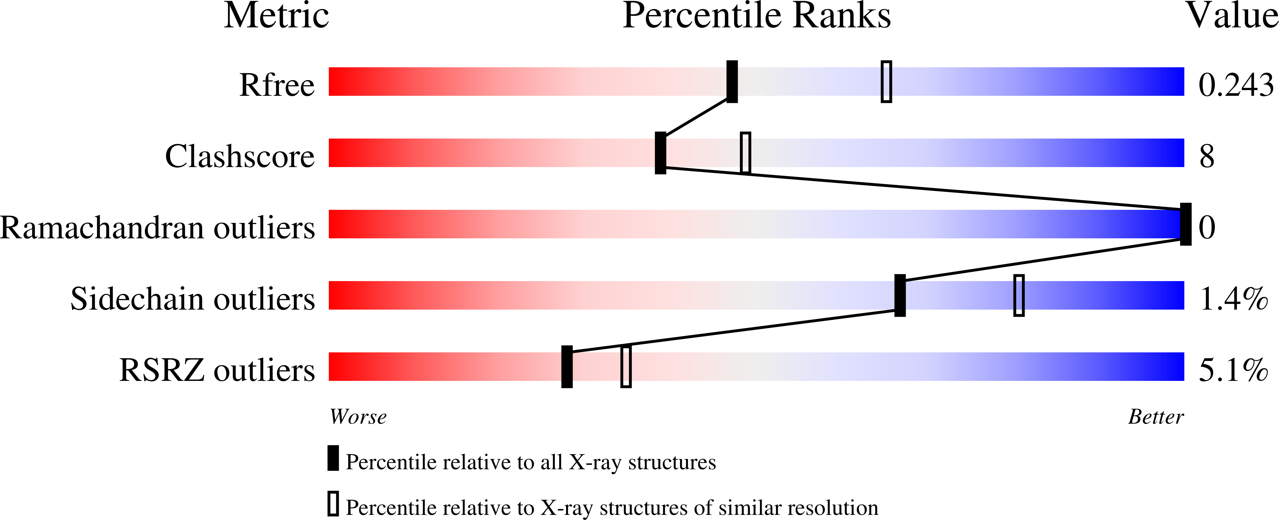
Resolution:
2.30 Å
R-Value Free:
0.25
R-Value Work:
0.19
R-Value Observed:
0.19
Space Group:
P 1 21 1
Macromolecular Entities
Structures with similar UniProt ID
Protein Blast
Polymer Type:polypeptide(L)
Molecule:DNA polymerase beta
Gene (Uniprot):POLB
Chain IDs:A
Chain Length:335
Number of Molecules:1
Biological Source:Homo sapiens
Modified Residue
Compound ID Chain ID Parent Comp ID Details 2D Image
8OG B DG ?
Primary Citation
Binary complex crystal structure of DNA polymerase beta reveals multiple conformations of the templating 8-oxoguanine lesion
Proc. Natl. Acad. Sci. U.S.A. 109 113 118 (2012)
PMID: 22178760 DOI: 10.1073/pnas.1112235108

Abstact

Oxidation of genomic DNA forms the guanine lesion 7,8-dihydro-8-oxoguanine (8-oxoG). When in the template base position during DNA synthesis the 8-oxoG lesion has dual coding potential by virtue of its anti- and syn-conformations, base pairing with cytosine and adenine, respectively. This impacts mutagenesis, because insertion of adenine opposite template 8-oxoG can result in a G to T transversion. DNA polymerases vary by orders of magnitude in their preferences for mutagenic vs. error-free 8-oxoG lesion bypass. Yet, the structural basis for lesion bypass specificity is not well understood. The DNA base excision repair enzyme DNA polymerase (pol) β is presented with gap-filling synthesis opposite 8-oxoG during repair and has similar insertion efficiencies for dCTP and dATP. We report the structure of pol β in binary complex with template 8-oxoG in a base excision repair substrate. The structure reveals both the syn- and anti-conformations of template 8-oxoG in the confines of the polymerase active site, consistent with the dual coding observed kinetically for this enzyme. A ternary complex structure of pol β with the syn-8-oxoG:anti-A Hoogsteen base pair in the closed fully assembled preinsertion active site is also reported. The syn-conformation of 8-oxoG is stabilized by minor groove hydrogen bonding between the side chain of Arg283 and O8 of 8-oxoG. An adjustment in the position of the phosphodiester backbone 5'-phosphate enables 8-oxoG to adopt the syn-conformation.

Legend

Protein

Chemical

Disease

Primary Citation of related structures
Feedback Form
Name
Email
Institute
Feedback