Deposition Date
2011-02-09
Release Date
2011-04-06
Last Version Date
2023-09-13
Entry Detail
PDB ID:
3QO2
Keywords:
Title:
Structural insights for MPP8 chromodomain interaction with histone H3 lysine 9
Biological Source:
Source Organism(s):
Homo sapiens (Taxon ID: 9606)
synthetic construct (Taxon ID: 32630)
synthetic construct (Taxon ID: 32630)
Expression System(s):
Method Details:
Experimental Method:
Resolution:
2.49 Å
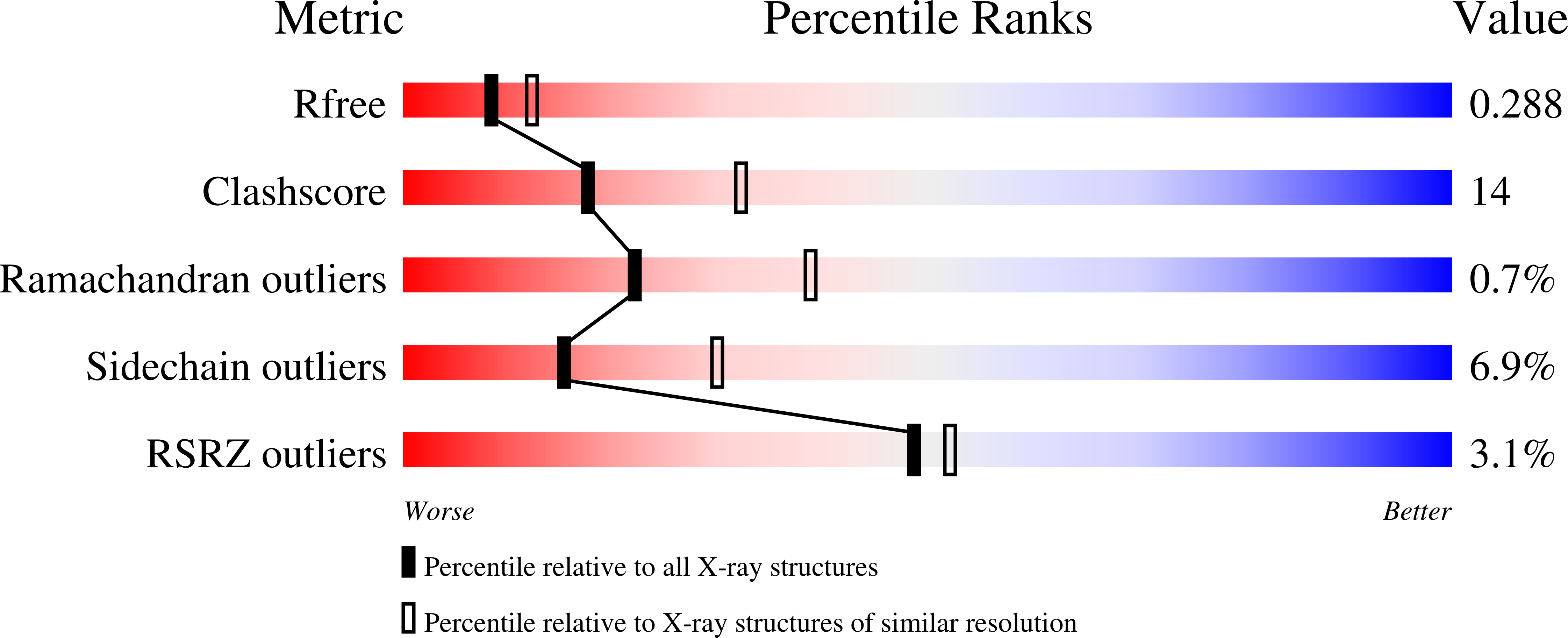
R-Value Free:
0.28
R-Value Work:
0.21
R-Value Observed:
0.21
Space Group:
P 43 21 2