3PE7 image
Deposition Date 2010-10-25
Release Date 2010-11-03
Last Version Date 2023-09-06
Entry Detail
PDB ID:
3PE7
Keywords:
Title:
Oligogalacturonate lyase in complex with manganese
Biological Source:
Expression System(s):
Method Details:
Experimental Method:
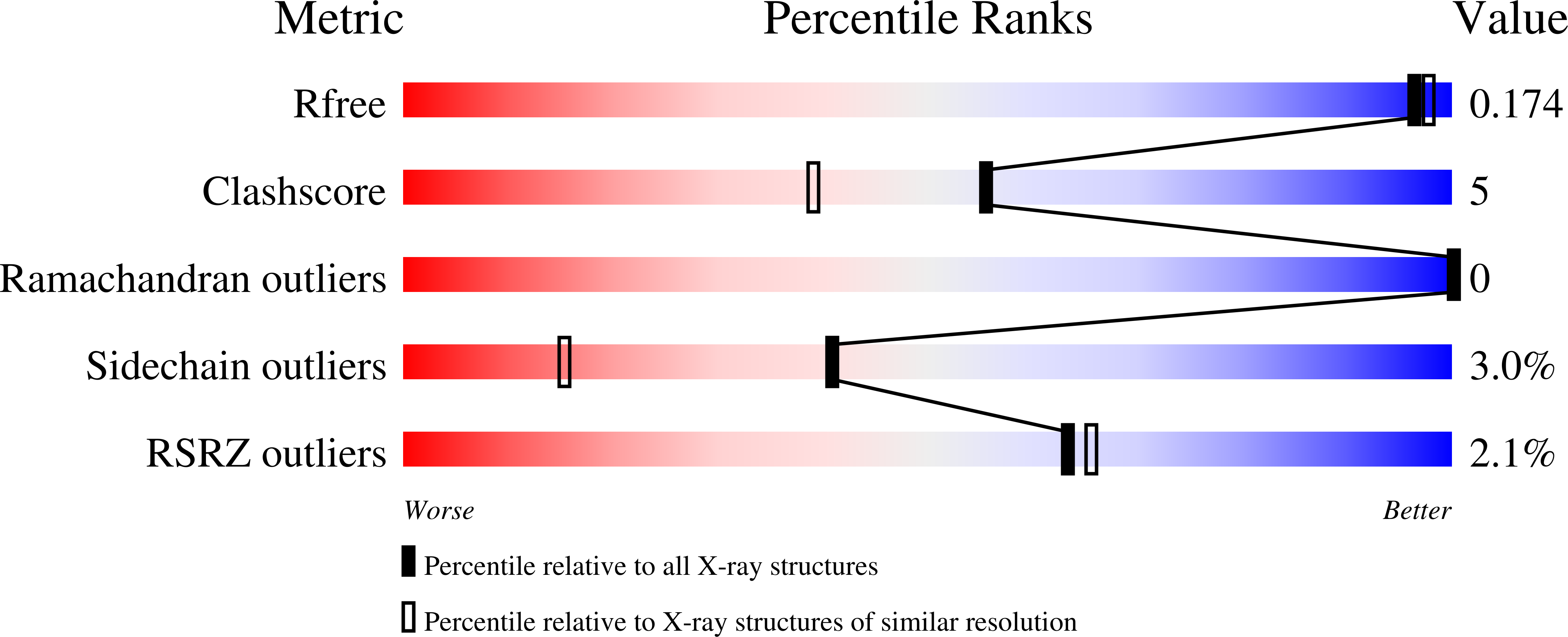
Resolution:
1.65 Å
R-Value Free:
0.20
R-Value Work:
0.17
R-Value Observed:
0.17
Space Group:
P 21 21 21
Macromolecular Entities
Structures with similar UniProt ID
Protein Blast
Polymer Type:polypeptide(L)
Molecule:Oligogalacturonate lyase
Gene (Uniprot):ogl
Chain IDs:A
Chain Length:388
Number of Molecules:1
Biological Source:Yersinia enterocolitica subsp. enterocolitica
Primary Citation
The active site of oligogalacturonate lyase provides unique insights into cytoplasmic oligogalacturonate beta-elimination.
J. Biol. Chem. 285 39029 39038 (2010)
PMID: 20851883 DOI: 10.1074/jbc.M110.153981

Abstact

Oligogalacturonate lyases (OGLs; now also classified as pectate lyase family 22) are cytoplasmic enzymes found in pectinolytic members of Enterobacteriaceae, such as the enteropathogen Yersinia enterocolitica. OGLs utilize a β-elimination mechanism to preferentially catalyze the conversion of saturated and unsaturated digalacturonate into monogalacturonate and the 4,5-unsaturated monogalacturonate-like molecule, 5-keto-4-deoxyuronate. To provide mechanistic insights into the specificity of this enzyme activity, we have characterized the OGL from Y. enterocolitica, YeOGL, on oligogalacturonides and determined its three-dimensional x-ray structure to 1.65 Å. The model contains a Mn(2+) atom in the active site, which is coordinated by three histidines, one glutamine, and an acetate ion. The acetate mimics the binding of the uronate group of galactourono-configured substrates. These findings, in combination with enzyme kinetics and metal supplementation assays, provide a framework for modeling the active site architecture of OGL. This enzyme appears to contain a histidine for the abstraction of the α-proton in the -1 subsite, a residue that is highly conserved throughout the OGL family and represents a unique catalytic base among pectic active lyases. In addition, we present a hypothesis for an emerging relationship observed between the cellular distribution of pectate lyase folding and the distinct metal coordination chemistries of pectate lyases.

Legend

Protein

Chemical

Disease

Primary Citation of related structures
Feedback Form
Name
Email
Institute
Feedback