Deposition Date
2010-09-21
Release Date
2010-11-10
Last Version Date
2023-09-06
Entry Detail
PDB ID:
3OXC
Keywords:
Title:
Wild Type HIV-1 Protease with Antiviral Drug Saquinavir
Biological Source:
Source Organism(s):
Human immunodeficiency virus 1 (Taxon ID: 11676)
Expression System(s):
Method Details:
Experimental Method:
Resolution:
1.16 Å
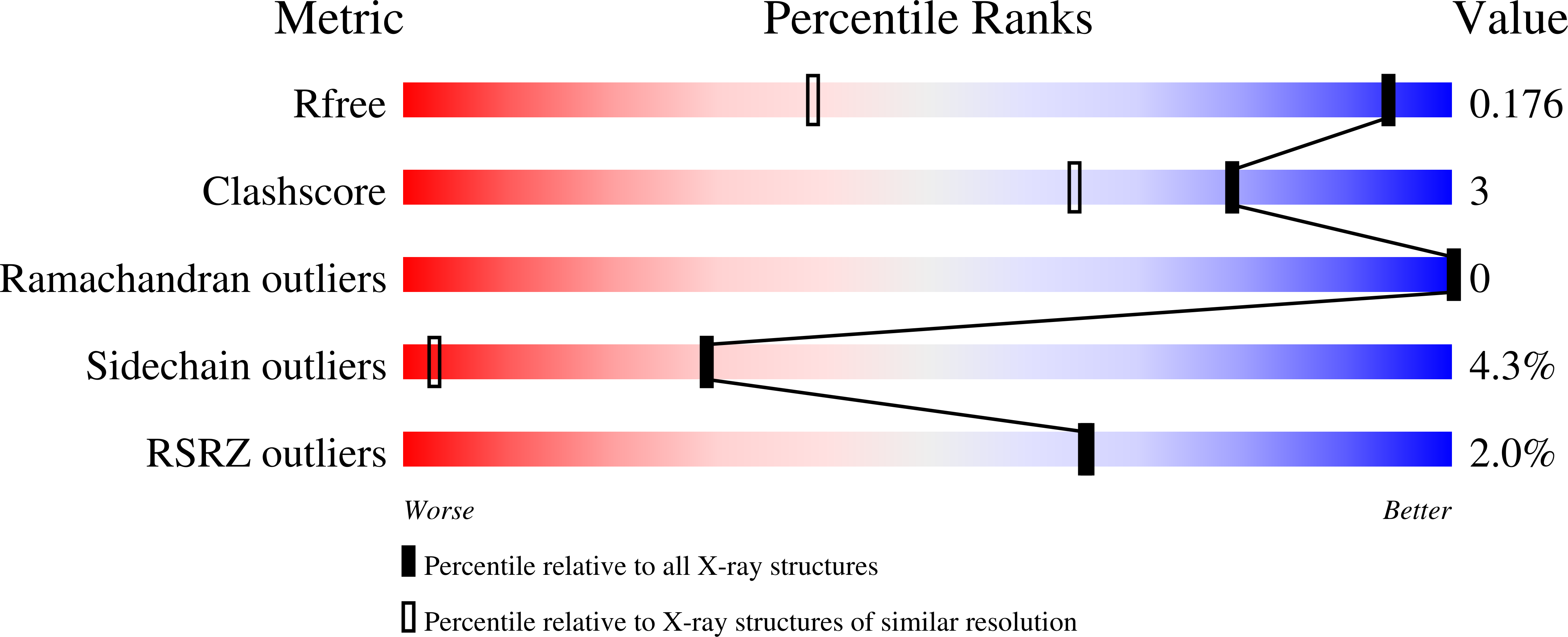
R-Value Free:
0.17
R-Value Work:
0.13
R-Value Observed:
0.13
Space Group:
P 21 21 21