3FWP image
Deposition Date 2009-01-19
Release Date 2010-01-19
Last Version Date 2023-09-06
Entry Detail
PDB ID:
3FWP
Keywords:
Title:
X-ray structure of uridine nucleoside phosphorylease from Salmonella typhimurium complexed with phosphate and its inhibitor 2,2'-anhydrouridine at 1.86 A resolution
Biological Source:
Source Organism(s):
Expression System(s):
Method Details:
Experimental Method:
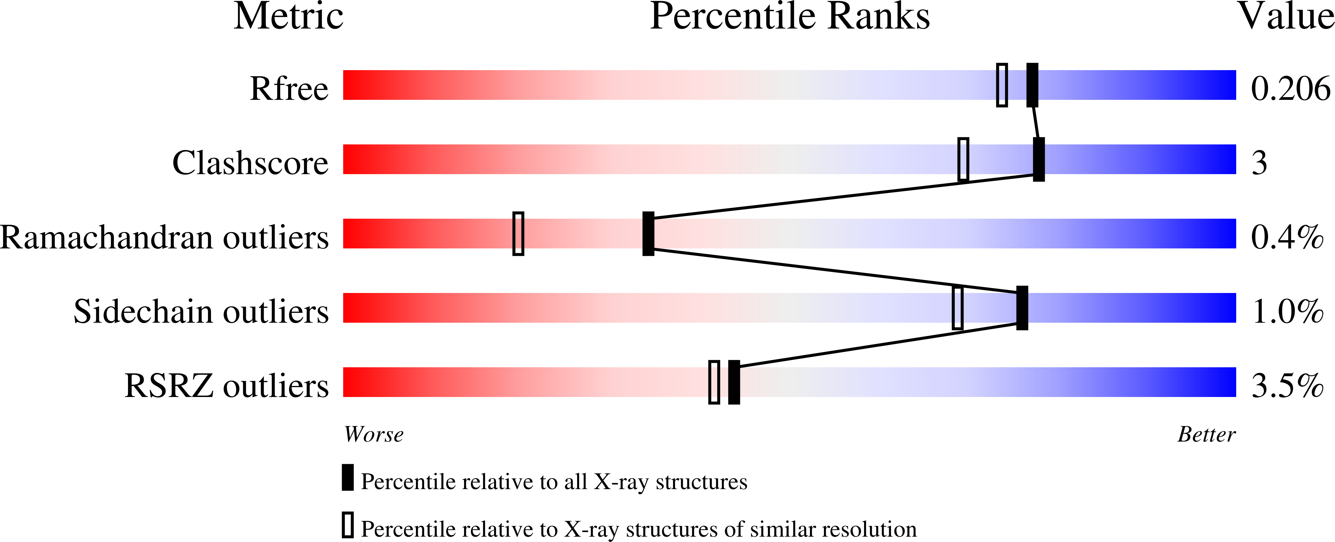
Resolution:
1.86 Å
R-Value Free:
0.20
R-Value Work:
0.17
R-Value Observed:
0.17
Space Group:
P 21 21 21
Macromolecular Entities
Structures with similar UniProt ID
Protein Blast
Polymer Type:polypeptide(L)
Molecule:Uridine phosphorylase
Gene (Uniprot):udp
Chain IDs:A, B, C, D, E, F
Chain Length:253
Number of Molecules:6
Biological Source:Salmonella typhimurium
Primary Citation
The X-ray structure of Salmonella typhimurium uridine nucleoside phosphorylase complexed with 2,2'-anhydrouridine, phosphate and potassium ions at 1.86 A resolution.
Acta Crystallogr. D Biol. Crystallogr. 66 51 60 (2010)
PMID: 20057049 DOI: 10.1107/S0907444909044175

Abstact

Uridine nucleoside phosphorylase is an important drug target for the development of anti-infective and antitumour agents. The X-ray crystal structure of Salmonella typhimurium uridine nucleoside phosphorylase (StUPh) complexed with its inhibitor 2,2'-anhydrouridine, phosphate and potassium ions has been solved and refined at 1.86 A resolution (R(cryst) = 17.6%, R(free) = 20.6%). The complex of human uridine phosphorylase I (HUPhI) with 2,2'-anhydrouridine was modelled using a computational approach. The model allowed the identification of atomic groups in 2,2'-anhydrouridine that might improve the interaction of future inhibitors with StUPh and HUPhI.

Legend

Protein

Chemical

Disease

Primary Citation of related structures
Feedback Form
Name
Email
Institute
Feedback