Deposition Date
2009-01-15
Release Date
2009-01-27
Last Version Date
2023-09-06
Entry Detail
PDB ID:
3FVE
Keywords:
Title:
Crystal structure of diaminopimelate epimerase Mycobacterium tuberculosis DapF
Biological Source:
Source Organism(s):
Mycobacterium tuberculosis (Taxon ID: 1773)
Expression System(s):
Method Details:
Experimental Method:
Resolution:
2.60 Å
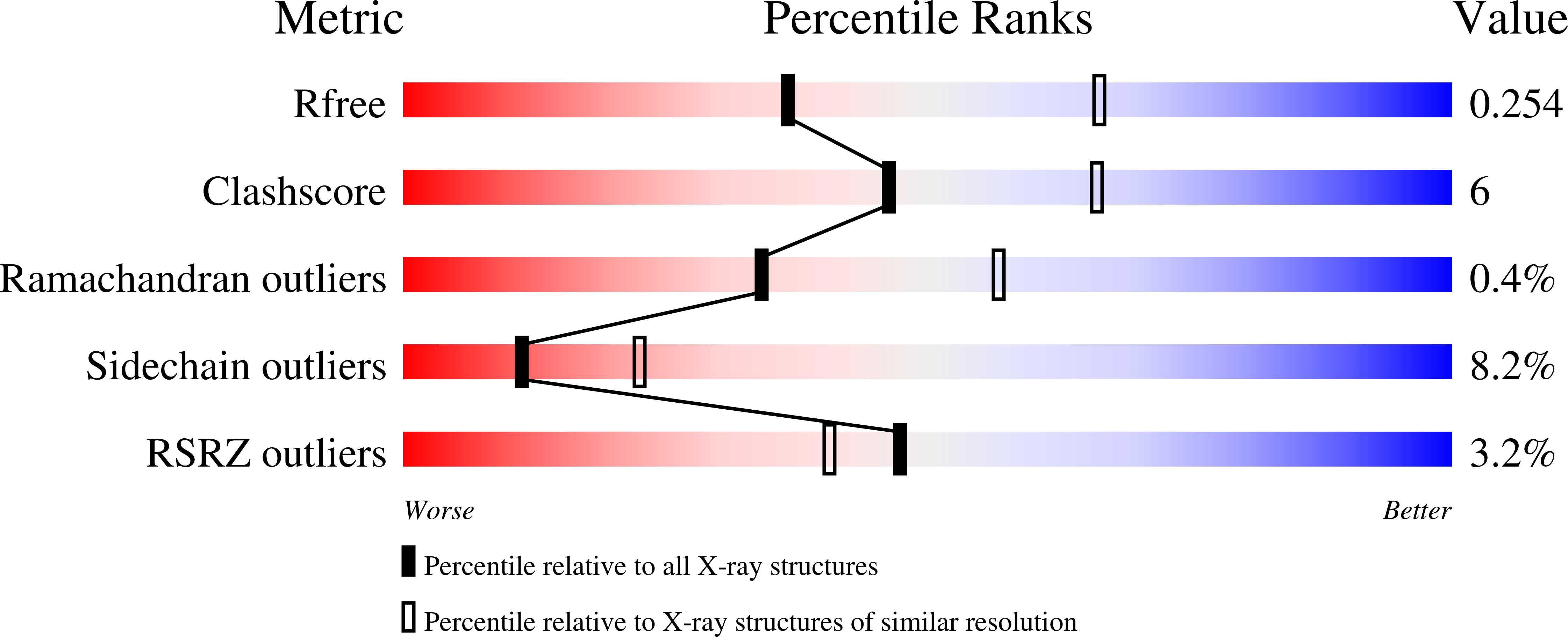
R-Value Free:
0.25
R-Value Work:
0.22
R-Value Observed:
0.22
Space Group:
P 65 2 2