

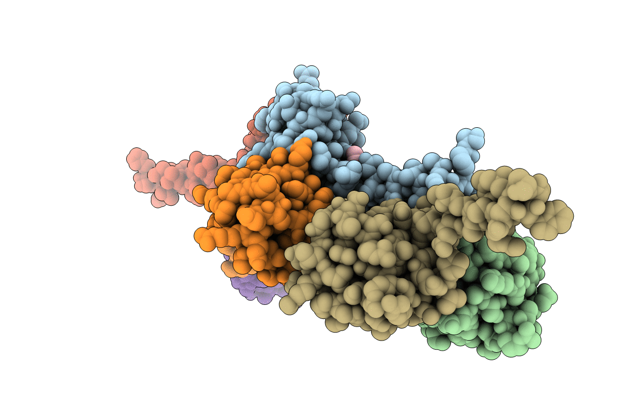
3F51 image
Deposition Date 2008-11-03
Release Date 2008-11-18
Last Version Date 2023-12-27
Entry Detail
PDB ID:
3F51
Title:
Crystal Structure of the clp gene regulator ClgR from Corynebacterium glutamicum
Biological Source:
Source Organism(s):
Expression System(s):
Method Details:
Experimental Method:
Resolution:
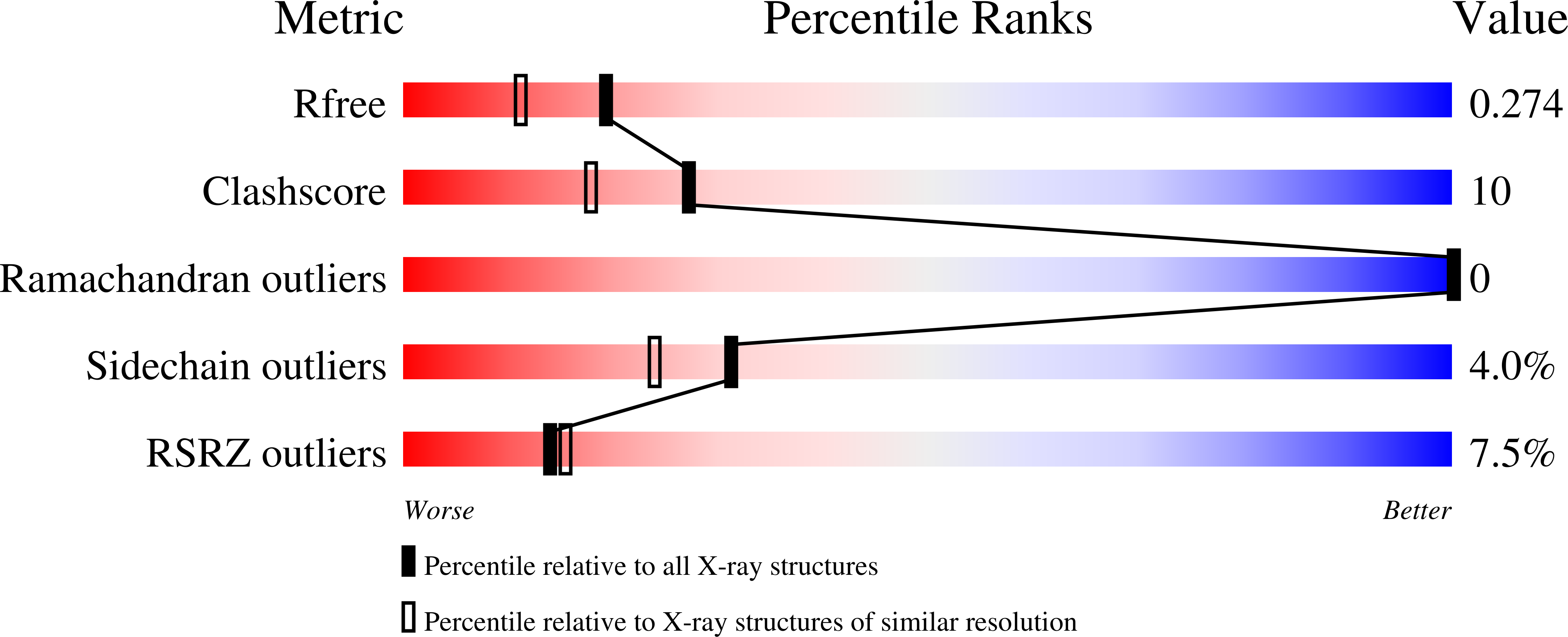
2.05 Å
R-Value Free:
0.23
R-Value Work:
0.21
R-Value Observed:
0.21
Space Group:
P 1 21 1
Macromolecular Entities
Structures with similar UniProt ID
Protein Blast
Polymer Type:polypeptide(L)
Molecule:Clp gene regulator (ClgR)
Gene (Uniprot):Cgl1962
Chain IDs:A, B, C, D, E, F
Chain Length:117
Number of Molecules:6
Biological Source:Corynebacterium glutamicum
Primary Citation
Crystal structure of the caseinolytic protease gene regulator, a transcriptional activator in actinomycetes
J. Biol. Chem. 284 5208 5216 (2009)
PMID: 19019826 DOI: 10.1074/jbc.M806591200

Abstact

Human pathogens of the genera Corynebacterium and Mycobacterium possess the transcriptional activator ClgR (clp gene regulator) which in Corynebacterium glutamicum has been shown to regulate the expression of the ClpCP protease genes. ClgR specifically binds to pseudo-palindromic operator regions upstream of clpC and clpP1P2. Here, we present the first crystal structure of a ClgR protein from C. glutamicum. The structure was determined from two different crystal forms to resolutions of 1.75 and 2.05 A, respectively. ClgR folds into a five-helix bundle with a helix-turn-helix motif typical for DNA-binding proteins. Upon dimerization the two DNA-recognition helices are arranged opposite to each other at the protein surface in a distance of approximately 30 A, which suggests that they bind into two adjacent major grooves of B-DNA in an anti-parallel manner. A binding pocket is situated at a strategic position in the dimer interface and could possess a regulatory role altering the positions of the DNA-binding helices.

Legend

Protein

Chemical

Disease

Primary Citation of related structures
Feedback Form
Name
Email
Institute
Feedback