3EY9 image
Deposition Date 2008-10-20
Release Date 2008-11-04
Last Version Date 2023-11-01
Entry Detail
PDB ID:
3EY9
Keywords:
Title:
Structural basis for membrane binding and catalytic activation of the peripheral membrane enzyme pyruvate oxidase from Escherichia coli
Biological Source:
Source Organism(s):
Escherichia coli (Taxon ID: 562)
Expression System(s):
Method Details:
Experimental Method:
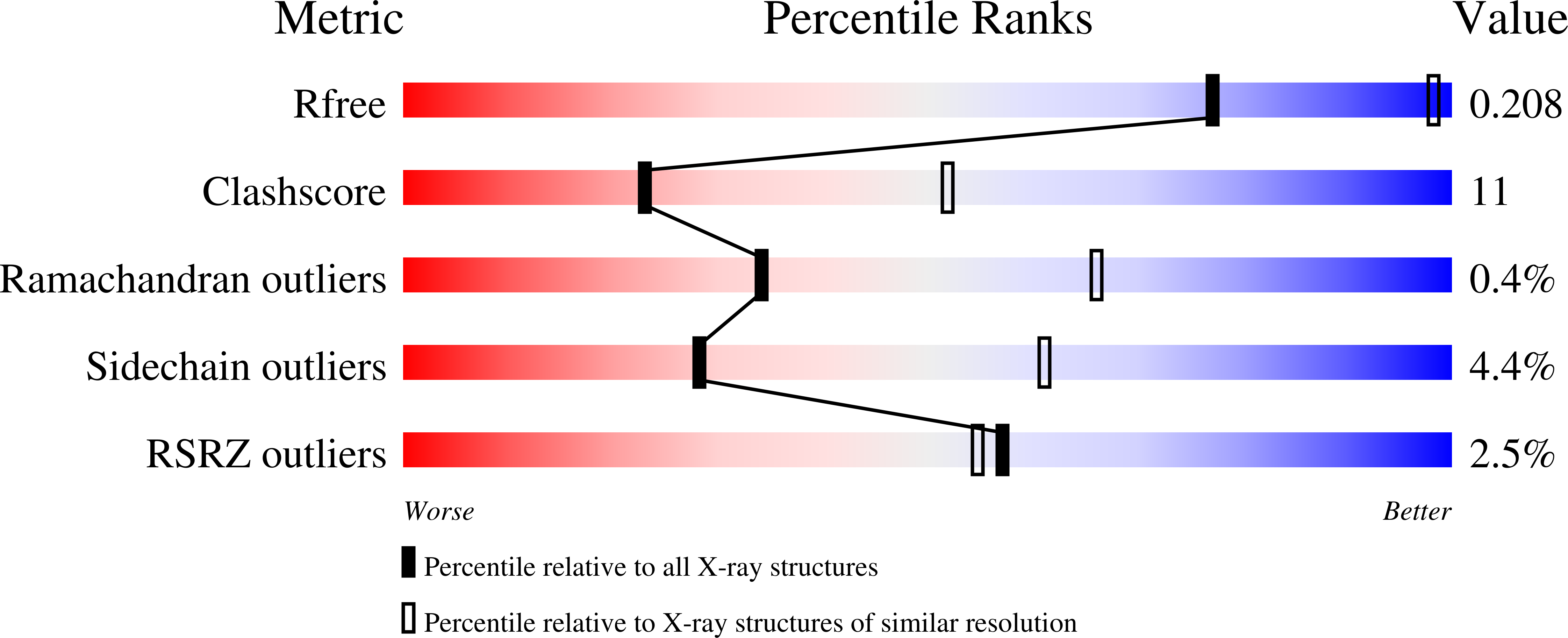
Resolution:
2.90 Å
R-Value Free:
0.21
R-Value Work:
0.18
R-Value Observed:
0.18
Space Group:
P 43 21 2
Macromolecular Entities
Structures with similar UniProt ID
Protein Blast
Polymer Type:polypeptide(L)
Molecule:Pyruvate dehydrogenase [cytoc
Gene (Uniprot):poxB
Chain IDs:A, B
Chain Length:572
Number of Molecules:2
Biological Source:Escherichia coli
Primary Citation
Structural basis for membrane binding and catalytic activation of the peripheral membrane enzyme pyruvate oxidase from Escherichia coli.
Proc. Natl. Acad. Sci. U.S.A. 105 17390 17395 (2008)
PMID: 18988747 DOI: 10.1073/pnas.0805027105

Abstact

The thiamin- and flavin-dependent peripheral membrane enzyme pyruvate oxidase from E. coli catalyzes the oxidative decarboxylation of the central metabolite pyruvate to CO(2) and acetate. Concomitant reduction of the enzyme-bound flavin triggers membrane binding of the C terminus and shuttling of 2 electrons to ubiquinone 8, a membrane-bound mobile carrier of the electron transport chain. Binding to the membrane in vivo or limited proteolysis in vitro stimulate the catalytic proficiency by 2 orders of magnitude. The molecular mechanisms by which membrane binding and activation are governed have remained enigmatic. Here, we present the X-ray crystal structures of the full-length enzyme and a proteolytically activated truncation variant lacking the last 23 C-terminal residues inferred as important in membrane binding. In conjunction with spectroscopic results, the structural data pinpoint a conformational rearrangement upon activation that exposes the autoinhibitory C terminus, thereby freeing the active site. In the activated enzyme, Phe-465 swings into the active site and wires both cofactors for efficient electron transfer. The isolated C terminus, which has no intrinsic helix propensity, folds into a helical structure in the presence of micelles.

Legend

Protein

Chemical

Disease

Primary Citation of related structures
Feedback Form
Name
Email
Institute
Feedback