3DY4 image
Deposition Date 2008-07-25
Release Date 2008-11-04
Last Version Date 2024-10-30
Entry Detail
PDB ID:
3DY4
Keywords:
Title:
Crystal structure of yeast 20S proteasome in complex with spirolactacystin
Biological Source:
Source Organism(s):
Method Details:
Experimental Method:
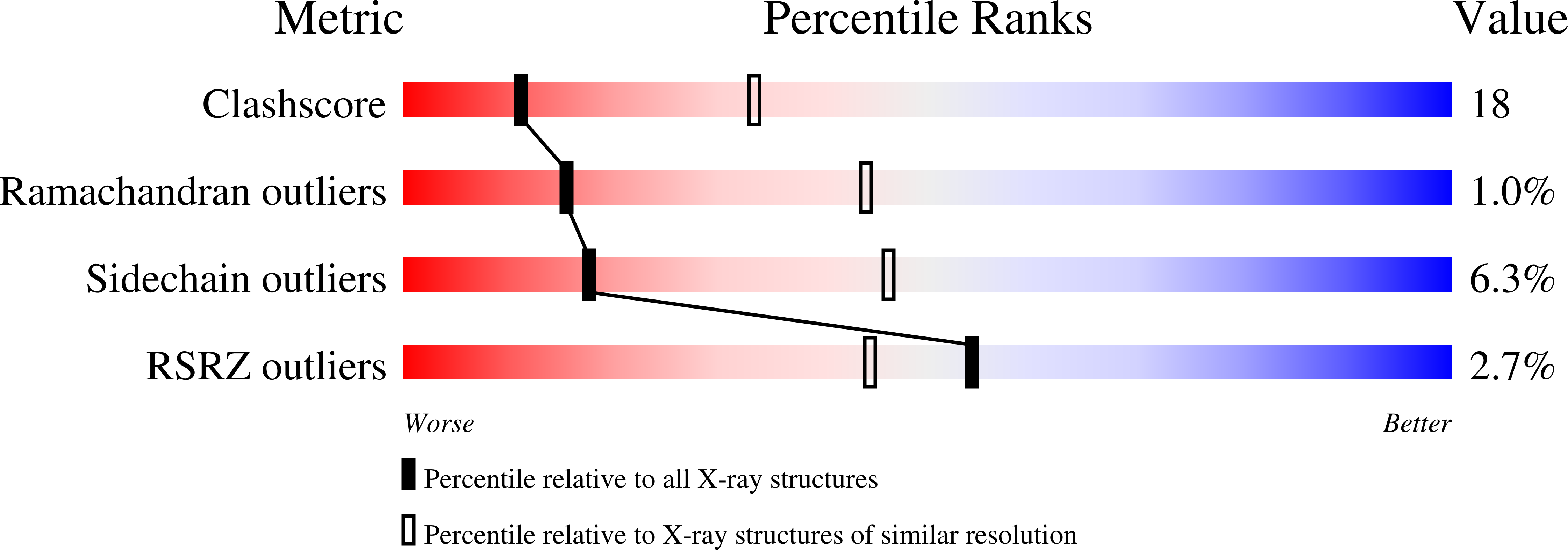
Resolution:
2.80 Å
R-Value Free:
0.25
R-Value Work:
0.22
R-Value Observed:
0.22
Space Group:
P 1 21 1
Macromolecular Entities
Structures with similar UniProt ID
Protein Blast
Polymer Type:polypeptide(L)
Molecule:Proteasome component Y7
Gene (Uniprot):PRE8
Chain IDs:A, O
Chain Length:250
Number of Molecules:2
Biological Source:Saccharomyces cerevisiae
Structures with similar UniProt ID
Protein Blast
Polymer Type:polypeptide(L)
Molecule:Proteasome component Y13
Gene (Uniprot):PRE9
Chain IDs:B, P
Chain Length:244
Number of Molecules:2
Biological Source:Saccharomyces cerevisiae
Structures with similar UniProt ID
Protein Blast
Polymer Type:polypeptide(L)
Molecule:Proteasome component PRE6
Gene (Uniprot):PRE6
Chain IDs:C, Q
Chain Length:241
Number of Molecules:2
Biological Source:Saccharomyces cerevisiae
Structures with similar UniProt ID
Protein Blast
Polymer Type:polypeptide(L)
Molecule:Proteasome component PUP2
Gene (Uniprot):PUP2
Chain IDs:D, R
Chain Length:242
Number of Molecules:2
Biological Source:Saccharomyces cerevisiae
Structures with similar UniProt ID
Protein Blast
Polymer Type:polypeptide(L)
Molecule:Proteasome component PRE5
Gene (Uniprot):PRE5
Chain IDs:E, S
Chain Length:233
Number of Molecules:2
Biological Source:Saccharomyces cerevisiae
Structures with similar UniProt ID
Protein Blast
Polymer Type:polypeptide(L)
Molecule:Proteasome component C1
Gene (Uniprot):PRE10
Chain IDs:F, T
Chain Length:244
Number of Molecules:2
Biological Source:Saccharomyces cerevisiae
Structures with similar UniProt ID
Protein Blast
Polymer Type:polypeptide(L)
Molecule:Proteasome component C7-alpha
Gene (Uniprot):SCL1
Chain IDs:G, U
Chain Length:243
Number of Molecules:2
Biological Source:Saccharomyces cerevisiae
Structures with similar UniProt ID
Protein Blast
Polymer Type:polypeptide(L)
Molecule:Proteasome component PUP1
Gene (Uniprot):PUP1
Chain IDs:H, V
Chain Length:222
Number of Molecules:2
Biological Source:Saccharomyces cerevisiae
Structures with similar UniProt ID
Protein Blast
Polymer Type:polypeptide(L)
Molecule:Proteasome component PUP3
Gene (Uniprot):PUP3
Chain IDs:I, W
Chain Length:204
Number of Molecules:2
Biological Source:Saccharomyces cerevisiae
Structures with similar UniProt ID
Protein Blast
Polymer Type:polypeptide(L)
Molecule:Proteasome component C11
Gene (Uniprot):PRE1
Chain IDs:J, X
Chain Length:198
Number of Molecules:2
Biological Source:Saccharomyces cerevisiae
Structures with similar UniProt ID
Protein Blast
Polymer Type:polypeptide(L)
Molecule:Proteasome component PRE2
Gene (Uniprot):PRE2
Chain IDs:K, Y
Chain Length:212
Number of Molecules:2
Biological Source:Saccharomyces cerevisiae
Structures with similar UniProt ID
Protein Blast
Polymer Type:polypeptide(L)
Molecule:Proteasome component C5
Gene (Uniprot):PRE7
Chain IDs:L, Z
Chain Length:222
Number of Molecules:2
Biological Source:Saccharomyces cerevisiae
Structures with similar UniProt ID
Protein Blast
Polymer Type:polypeptide(L)
Molecule:Proteasome component PRE4
Gene (Uniprot):PRE4
Chain IDs:M, AA (auth: 1)
Chain Length:233
Number of Molecules:2
Biological Source:Saccharomyces cerevisiae
Structures with similar UniProt ID
Protein Blast
Polymer Type:polypeptide(L)
Molecule:Proteasome component PRE3
Gene (Uniprot):PRE3
Chain IDs:N, BA (auth: 2)
Chain Length:196
Number of Molecules:2
Biological Source:Saccharomyces cerevisiae
Ligand Molecules
Primary Citation
Structural analysis of spiro beta-lactone proteasome inhibitors.
J.Am.Chem.Soc. 130 14981 14983 (2008)
PMID: 18928262 DOI: 10.1021/ja806059t

Abstact

Spiro beta-lactone-based proteasome inhibitors were discovered in the context of an asymmetric catalytic total synthesis of the natural product (+)-lactacystin (1). Lactone 4 was found to be a potent inhibitor of the 26S proteasome, while its C-6 epimer (5) displayed weak activity. Crystallographic studies of the two analogues covalently bound to the 20S proteasome permitted characterization of the important stabilizing interactions between each inhibitor and the proteasome's key catalytic N-terminal threonine residue. This structural data support the hypothesis that the discrepancy in potency between 4 and 5 may be due to differences in the hydrolytic stabilities of the resulting acyl enzyme complexes.

Legend

Protein

Chemical

Disease

Primary Citation of related structures
Feedback Form
Name
Email
Institute
Feedback