
Deposition Date
2008-05-19
Release Date
2009-05-19
Last Version Date
2023-11-01
Entry Detail
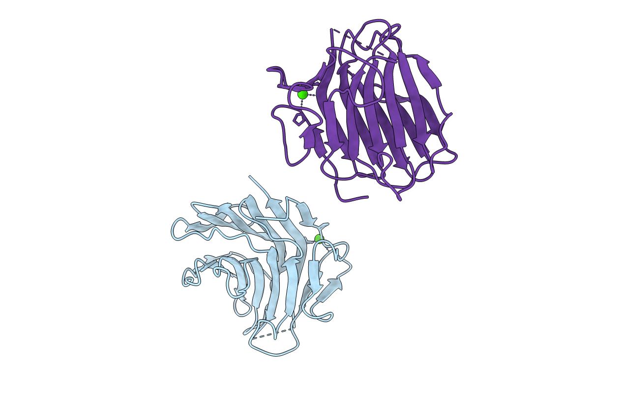
PDB ID:
3D6E
Keywords:
Title:
Crystal structure of the engineered 1,3-1,4-beta-glucanase protein from Bacillus licheniformis
Biological Source:
Source Organism(s):
Bacillus licheniformis (Taxon ID: 1402)
Expression System(s):
Method Details:
Experimental Method:
Resolution:
2.40 Å
R-Value Free:
0.25
R-Value Work:
0.21
R-Value Observed:
0.21
Space Group:
P 1
