2f2p image
Deposition Date 2005-11-17
Release Date 2006-02-21
Last Version Date 2023-08-23
Entry Detail
PDB ID:
2F2P
Title:
Structure of calmodulin bound to a calcineurin peptide: a new way of making an old binding mode
Biological Source:
Source Organism(s):
Bos taurus (Taxon ID: 9913)
Expression System(s):
Method Details:
Experimental Method:
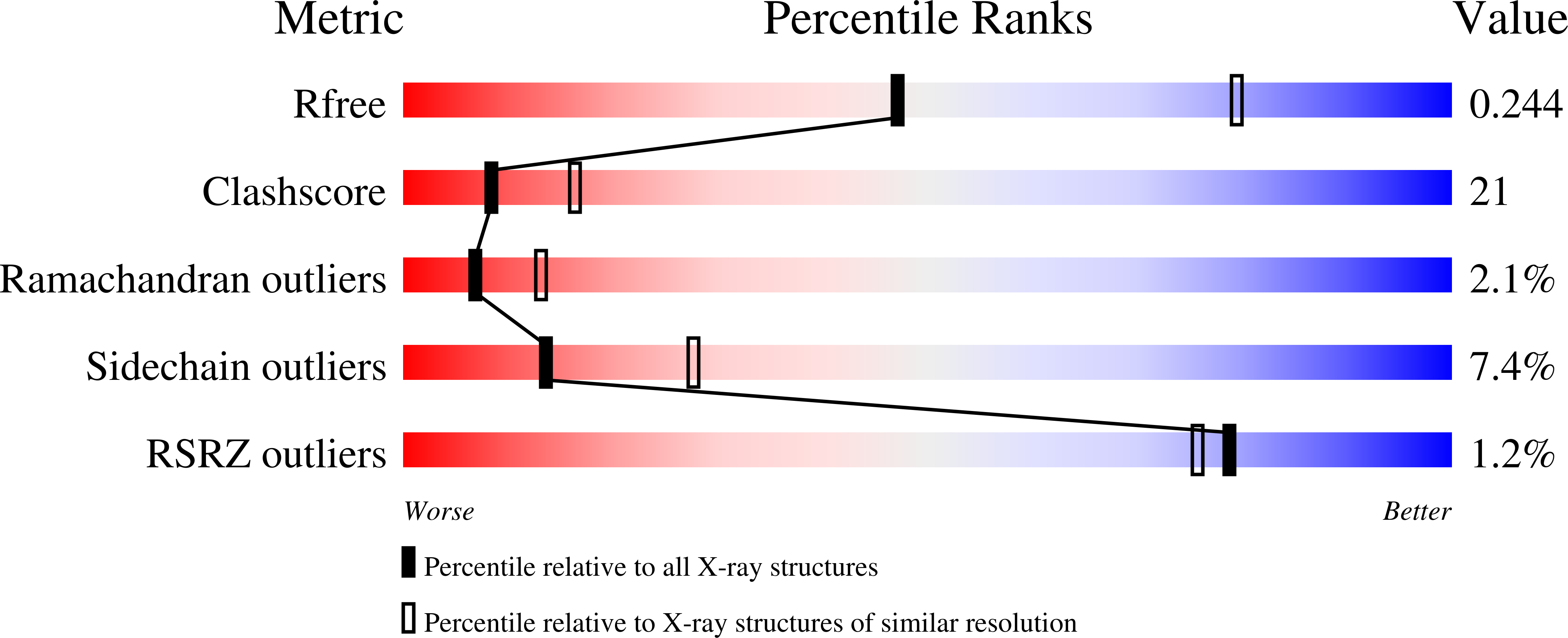
Resolution:
2.60 Å
R-Value Free:
0.29
R-Value Work:
0.23
R-Value Observed:
0.24
Space Group:
P 3 2 1
Macromolecular Entities
Protein Blast
Polymer Type:polypeptide(L)
Molecule:Calmodulin fused with calmodu
Gene (Uniprot):CALM
Chain IDs:A, B
Chain Length:179
Number of Molecules:2
Biological Source:Bos taurus
Ligand Molecules
Primary Citation
Structure of calmodulin bound to a calcineurin Peptide: a new way of making an old binding mode.
Biochemistry 45 738 745 (2006)
PMID: 16411749 DOI: 10.1021/bi0521801

Abstact

Calcineurin is a calmodulin-binding protein in brain and the only serine/threonine protein phosphatase under the control of Ca2+/calmodulin (CaM), which plays a critical role in coupling Ca2+ signals to cellular responses. CaM up-regulates the phosphatase activity of calcineurin by binding to the CaM-binding domain (CBD) of calcineurin subunit A. Here, we report crystal structural studies of CaM bound to a CBD peptide. The chimeric protein containing CaM and the CBD peptide forms an intimate homodimer, in which CaM displays a native-like extended conformation and the CBD peptide shows alpha-helical structure. Unexpectedly, the N-terminal lobe from one CaM and the C-terminal lobe from the second molecule form a combined binding site to trap the peptide. Thus, the dimer provides two binding sites, each of which is reminiscent of the fully collapsed conformation of CaM commonly observed in complex with, for example, the myosin light chain kinase (MLCK) peptide. The interaction between the peptide and CaM is highly specific and similar to MLCK.

Legend

Protein

Chemical

Disease

Primary Citation of related structures
Feedback Form
Name
Email
Institute
Feedback