

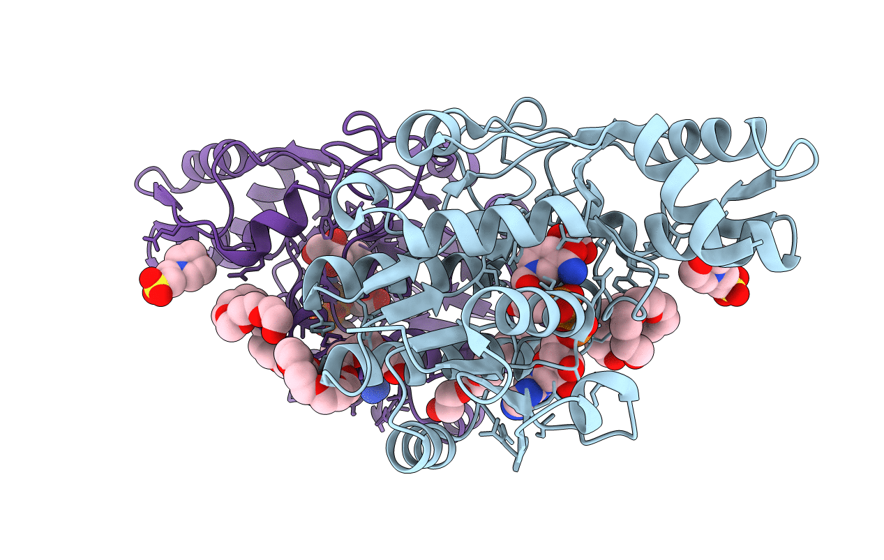
2YY7 image
Deposition Date 2007-04-27
Release Date 2008-04-29
Last Version Date 2024-03-13
Entry Detail
PDB ID:
2YY7
Keywords:
Title:
Crystal structure of thermolabile L-threonine dehydrogenase from Flavobacterium frigidimaris KUC-1
Biological Source:
Source Organism(s):
Method Details:
Experimental Method:
Resolution:
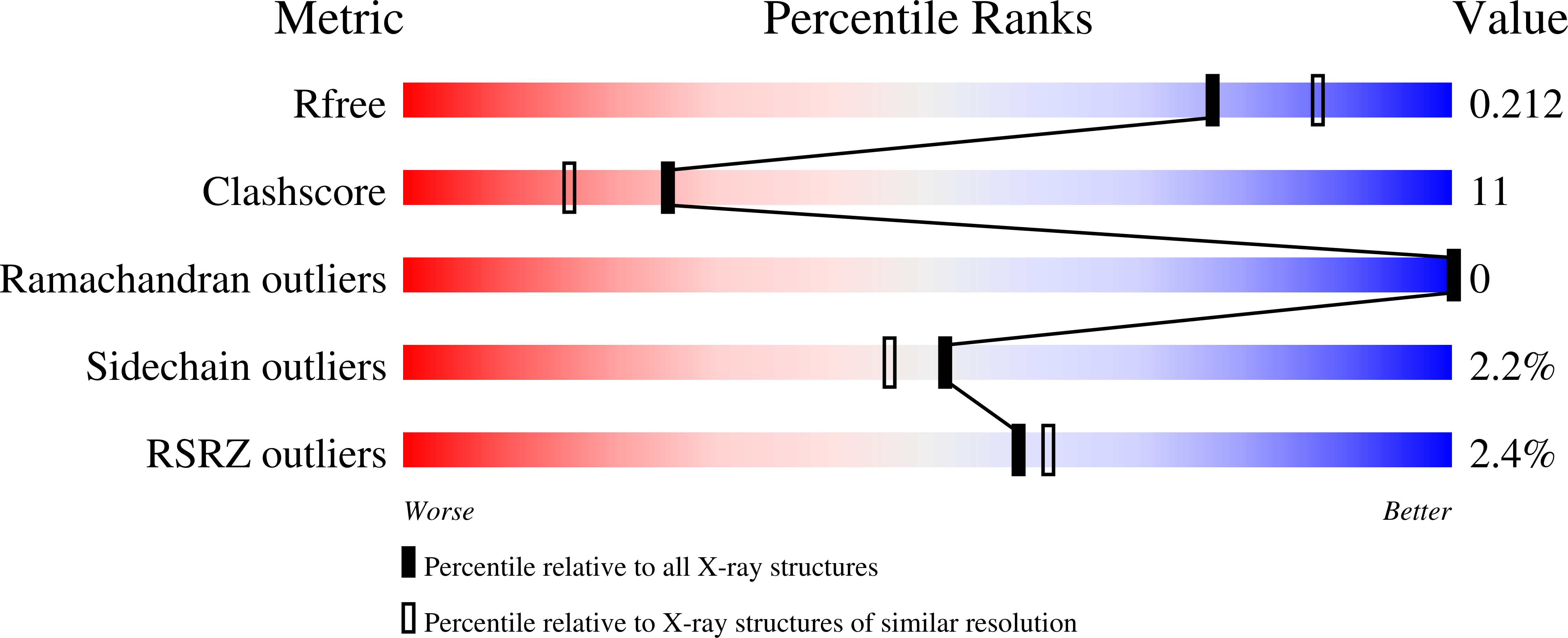
2.06 Å
R-Value Free:
0.20
R-Value Work:
0.19
Space Group:
P 21 21 21
Macromolecular Entities
Structures with similar UniProt ID
Protein Blast
Polymer Type:polypeptide(L)
Molecule:L-threonine dehydrogenase
Gene (Uniprot):ltd
Chain IDs:A, B
Chain Length:312
Number of Molecules:2
Biological Source:Flavobacterium frigidimaris
Primary Citation
Crystal structure of UDP-galactose 4-epimerase-like L-threonine dehydrogenase belonging to the intermediate short-chain dehydrogenase-reductase superfamily
FEBS J. 277 5124 5132 (2010)
PMID: 21078123 DOI: 10.1111/j.1742-4658.2010.07916.x

Abstact

The crystal structure of a L-threonine dehydrogenase (L-ThrDH; EC 1.1.1.103) from the psychrophilic bacterium Flavobacterium frigidimaris KUC-1, which shows no sequence similarity to conventional L-ThrDHs, was determined in the presence of NAD and a substrate analog, glycerol. The asymmetric unit consisted of two subunits related by a two-fold rotation axis. Each monomer consisted of a Rossmann-fold domain and a carboxyl-terminal catalytic domain. The overall fold of F. frigidimaris L-ThrDH showed significant similarity to that of UDP-galactose 4-epimerase (GalE); however, structural comparison of the enzyme with E. coli and human GalEs showed clear topological differences in three loops (loop 1, loop 2 and the NAD-binding loop) around the substrate and NAD binding sites. In F. frigidimaris L-ThrDH, loops 1 and 2 insert toward the active site cavity, creating a barrier preventing the binding of UDP-glucose. Alternatively, loop 1 contributes to a unique substrate binding pocket in the F. frigidimaris enzyme. The NAD binding loop, which tightly holds the adenine ribose moiety of NAD in the Escherichia coli and human GalEs, is absent in F. frigidimaris L-ThrDH. Consequently, the cofactor binds to F. frigidimaris L-ThrDH in a reversible manner, unlike its binding to GalE. The substrate binding model suggests that the reaction proceeds through abstraction of the β-hydroxyl hydrogen of L-threonine via either a proton shuttle mechanism driven by Tyr143 and facilitated by Ser118 or direct proton transfer driven by Tyr143. The present structure provides a clear bench mark for distinguishing GalE-like L-ThrDHs from GalEs.

Legend

Protein

Chemical

Disease

Primary Citation of related structures
Feedback Form
Name
Email
Institute
Feedback