2YID image
Deposition Date 2011-05-11
Release Date 2011-06-15
Last Version Date 2023-12-20
Entry Detail
PDB ID:
2YID
Keywords:
Title:
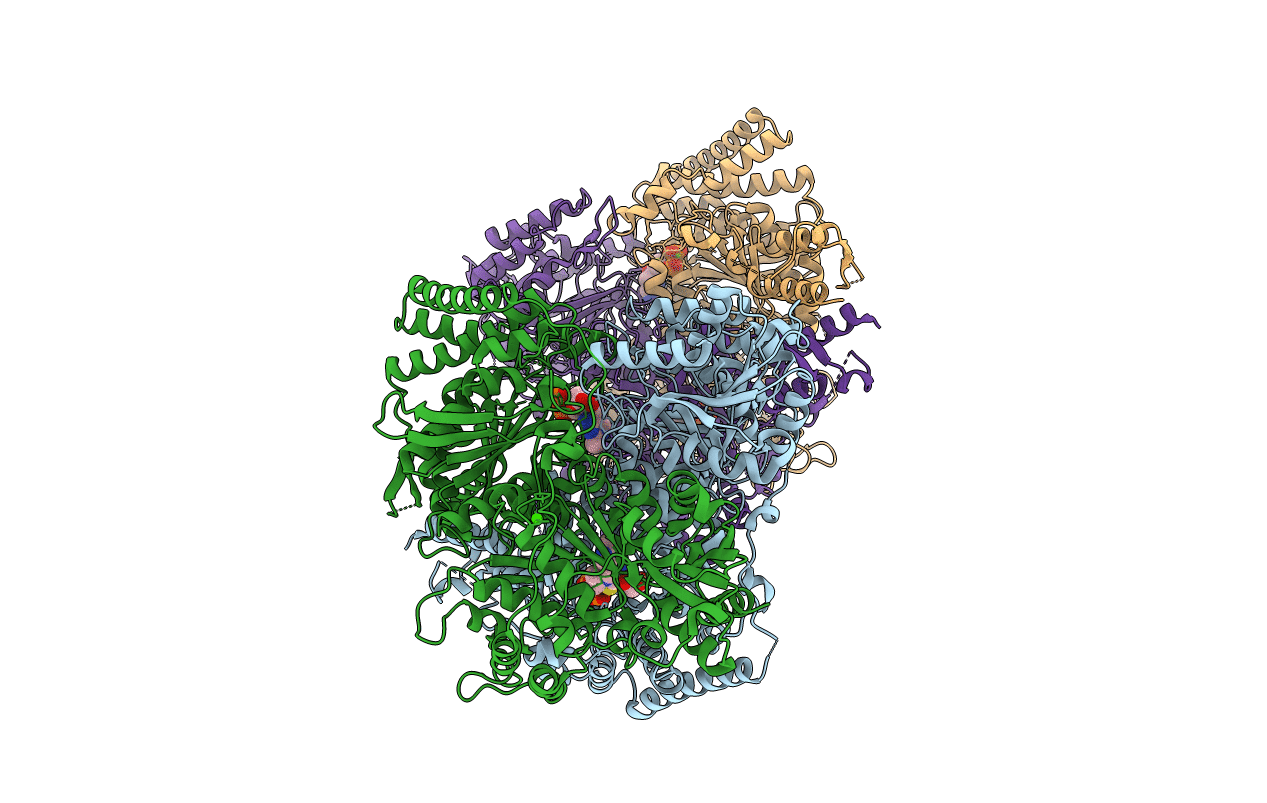
Crystal structure of the SucA domain of Mycobacterium smegmatis alpha- ketoglutarate decarboxylase in complex with the enamine-ThDP intermediate
Biological Source:
Source Organism(s):
Expression System(s):
Method Details:
Experimental Method:
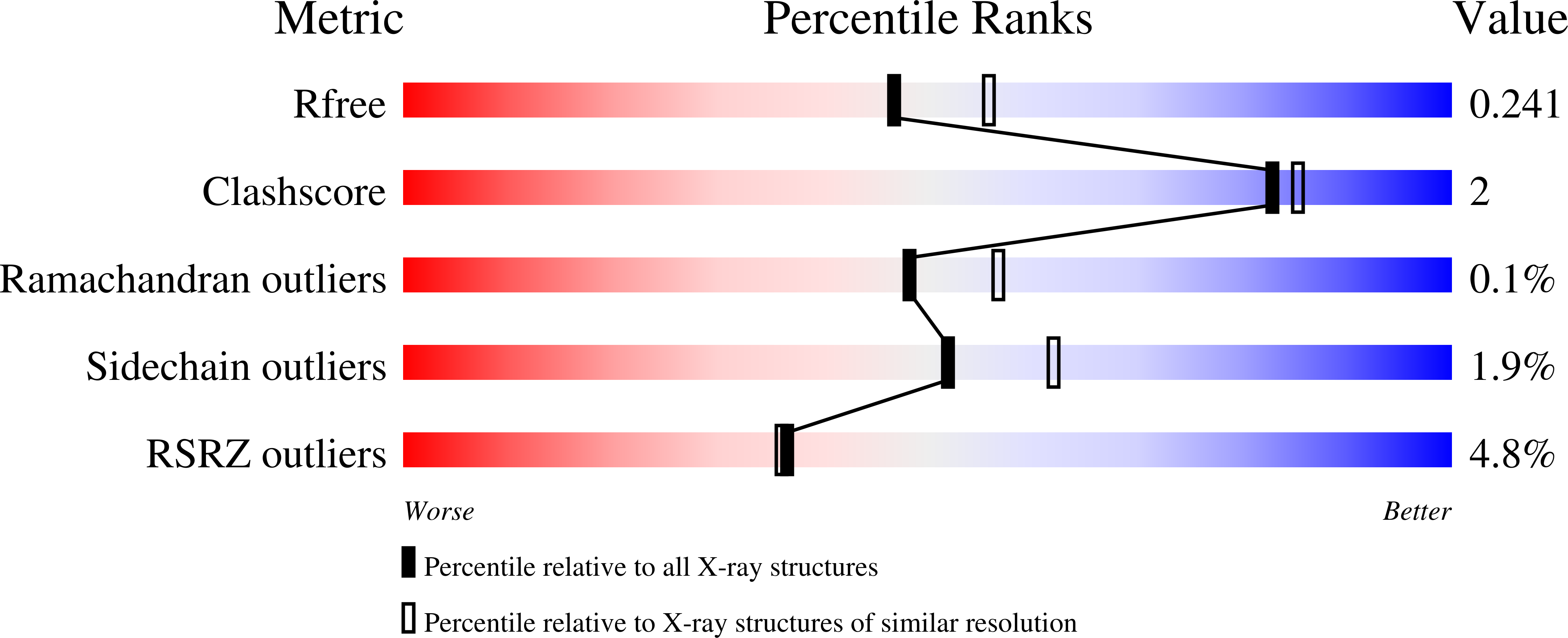
Resolution:
2.25 Å
R-Value Free:
0.22
R-Value Work:
0.19
R-Value Observed:
0.19
Space Group:
P 1
Macromolecular Entities
Structures with similar UniProt ID
Protein Blast
Polymer Type:polypeptide(L)
Molecule:2-OXOGLUTARATE DECARBOXYLASE
Gene (Uniprot):kgd
Chain IDs:A, B, C, D
Chain Length:868
Number of Molecules:4
Biological Source:MYCOBACTERIUM SMEGMATIS
Primary Citation
Functional Plasticity and Allosteric Regulation of Alpha-Ketoglutarate Decarboxylase in Central Mycobacterial Metabolism.
Chem. Biol. 18 1011 ? (2011)
PMID: 21867916 DOI: 10.1016/J.CHEMBIOL.2011.06.004

Abstact

The α-ketoglutarate dehydrogenase (KDH) complex is a major regulatory point of aerobic energy metabolism. Mycobacterium tuberculosis was reported to lack KDH activity, and the putative KDH E1o component, α-ketoglutarate decarboxylase (KGD), was instead assigned as a decarboxylase or carboligase. Here, we show that this protein does in fact sustain KDH activity, as well as the additional two reactions, and these multifunctional properties are shared by the Escherichia coli homolog, SucA. We also show that the mycobacterial enzyme is finely regulated by an additional acyltransferase-like domain and by the action of acetyl-CoA, a powerful allosteric activator able to enhance the concerted protein motions observed during catalysis. Our results uncover the functional plasticity of a crucial node in bacterial metabolism, which may be important for M. tuberculosis during host infection.

Legend

Protein

Chemical

Disease

Primary Citation of related structures
Feedback Form
Name
Email
Institute
Feedback