2NO2 image
Deposition Date 2006-10-24
Release Date 2007-10-30
Last Version Date 2023-12-27
Entry Detail
PDB ID:
2NO2
Keywords:
Title:
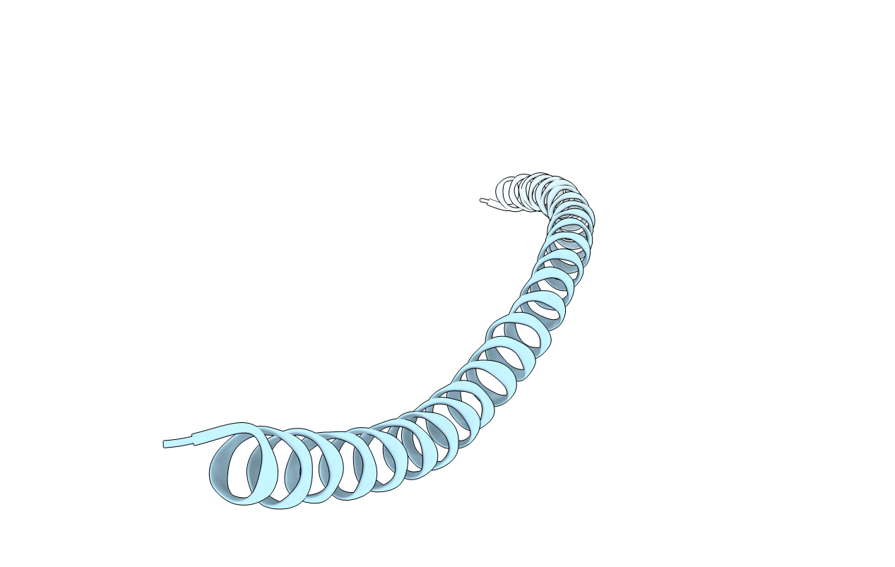
Crystal structure of the DLLRKN-containing coiled-coil domain of Huntingtin-interacting protein 1
Biological Source:
Source Organism(s):
Homo sapiens (Taxon ID: 9606)
Expression System(s):
Method Details:
Experimental Method:
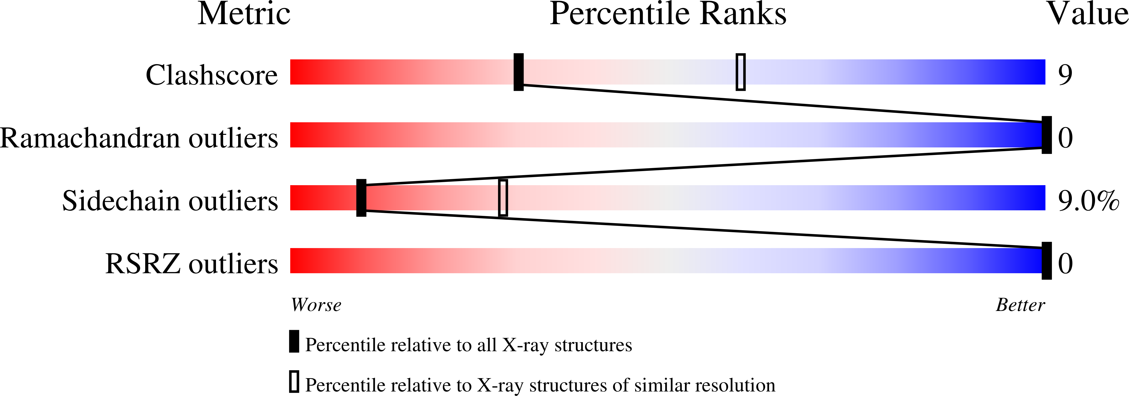
Resolution:
2.80 Å
R-Value Free:
0.31
R-Value Work:
0.28
R-Value Observed:
0.42
Space Group:
P 42 21 2
Macromolecular Entities
Structures with similar UniProt ID
Protein Blast
Polymer Type:polypeptide(L)
Molecule:Huntingtin-interacting protei
Gene (Uniprot):HIP1
Chain IDs:A
Chain Length:107
Number of Molecules:1
Biological Source:Homo sapiens
Primary Citation
Crystal structure at 2.8 A of the DLLRKN-containing coiled-coil domain of huntingtin-interacting protein 1 (HIP1) reveals a surface suitable for clathrin light chain binding
J. Mol. Biol. 367 8 15 (2007)
PMID: 17257618 DOI: 10.1016/j.jmb.2006.12.052

Abstact

Huntingtin interacting protein 1 (HIP1) is a member of a family of proteins whose interaction with Huntingtin is critical to prevent cells from initiating apoptosis. HIP1, and related protein HIP12/1R, can also bind to clathrin and membrane phospholipids, and HIP12/1R links the CCV to the actin cytoskeleton. HIP1 and HIP12/1R interact with the clathrin light chain EED regulatory site and stimulate clathrin lattice assembly. Here, we report the X-ray structure of the coiled-coil domain of HIP1 (residues 482-586) that includes residues crucial for binding clathrin light chain. The dimeric HIP1 crystal structure is partially splayed open. The comparison of the HIP1 model with coiled-coil predictions revealed the heptad repeat in the dimeric trunk (S2 path) is offset relative to the register of the heptad repeat from the N-terminal portion (S1 path) of the molecule. Furthermore, surface analysis showed there is a third hydrophobic path (S3) running parallel with S1 and S2. We present structural evidence supporting a role for the S3 path as an interaction surface for clathrin light chain. Finally, comparative analysis suggests the mode of binding between sla2p and clathrin light chain may be different in yeast.

Legend

Protein

Chemical

Disease

Primary Citation of related structures
Feedback Form
Name
Email
Institute
Feedback