Deposition Date
2006-10-23
Release Date
2006-11-07
Last Version Date
2024-10-09
Entry Detail
PDB ID:
2NMS
Keywords:
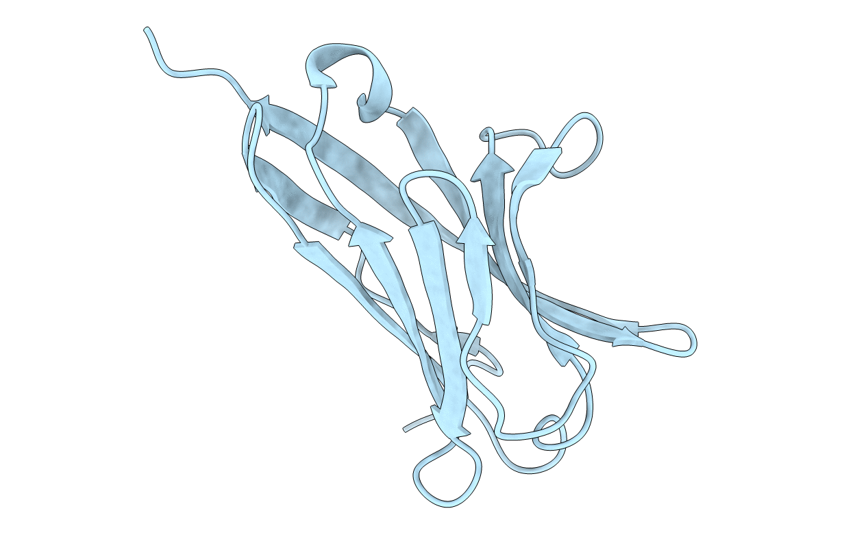
Title:
The Crystal Structure of the Extracellular Domain of the Inhibitor Receptor Expressed on Myeloid Cells IREM-1
Biological Source:
Source Organism(s):
Homo sapiens (Taxon ID: 9606)
Expression System(s):
Method Details:
Experimental Method:
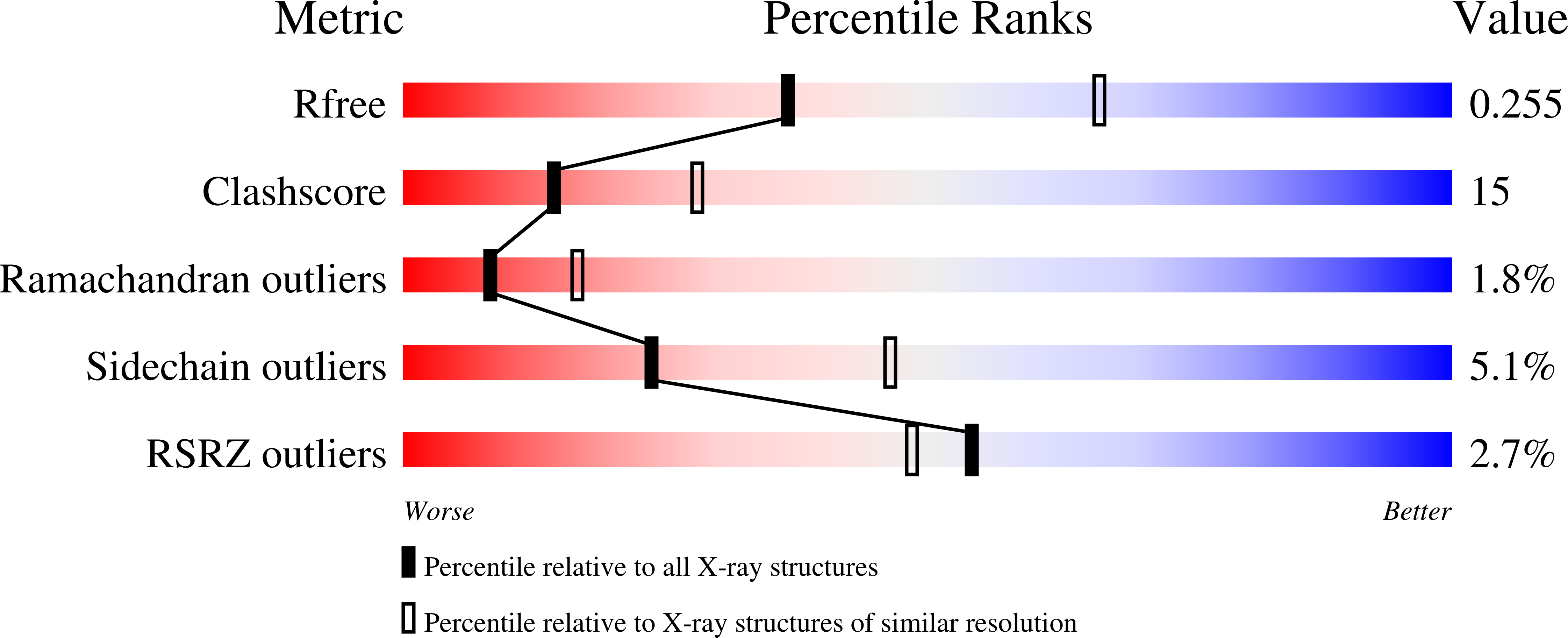
Resolution:
2.60 Å
R-Value Free:
0.25
R-Value Work:
0.21
Space Group:
P 31 2 1