2N2F image
Deposition Date 2015-05-06
Release Date 2015-09-09
Last Version Date 2024-05-15
Entry Detail
PDB ID:
2N2F
Title:
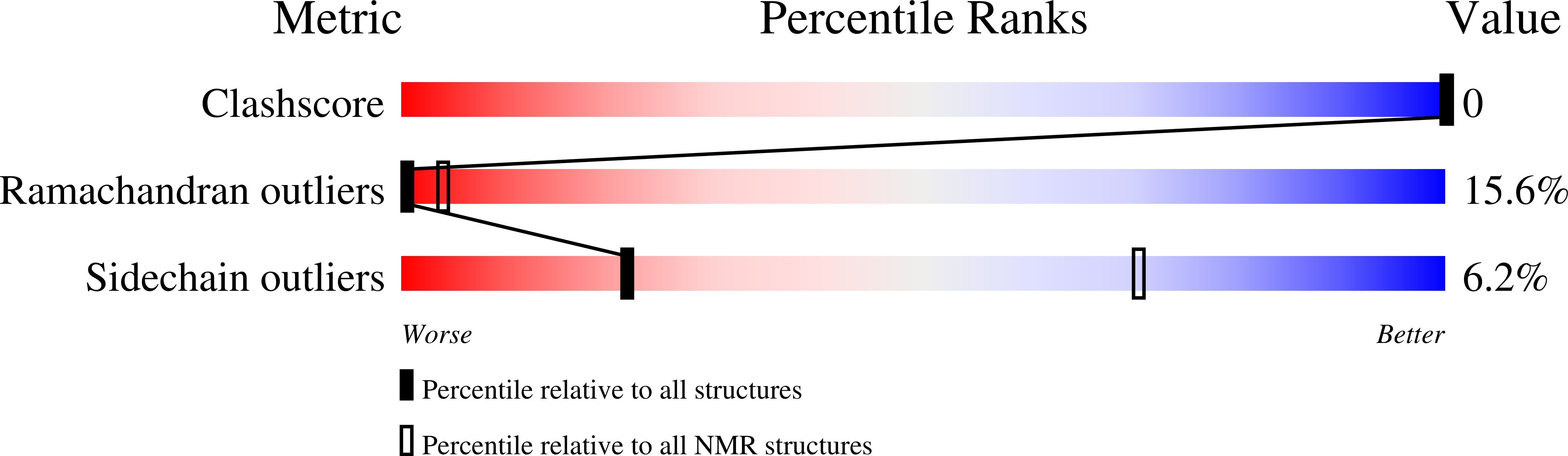
Solution NMR structure of Dynorphin 1-13 bound to Kappa Opioid Receptor
Biological Source:
Source Organism(s):
Homo sapiens (Taxon ID: 9606)
Method Details:
Experimental Method:
Conformers Calculated:
1000
Conformers Submitted:
10
Selection Criteria:
structures with the least restraint violations
Macromolecular Entities
Structures with similar UniProt ID
Protein Blast
Polymer Type:polypeptide(L)
Molecule:Dynorphin A(1-13)
Gene (Uniprot):PDYN
Chain IDs:A
Chain Length:13
Number of Molecules:1
Biological Source:Homo sapiens
Ligand Molecules
Primary Citation
NMR structure and dynamics of the agonist dynorphin peptide bound to the human kappa opioid receptor.
Proc. Natl. Acad. Sci. U.S.A. 112 11852 11857 (2015)
PMID: 26372966 DOI: 10.1073/pnas.1510117112

Abstact

The structure of the dynorphin (1-13) peptide (dynorphin) bound to the human kappa opioid receptor (KOR) has been determined by liquid-state NMR spectroscopy. (1)H and (15)N chemical shift variations indicated that free and bound peptide is in fast exchange in solutions containing 1 mM dynorphin and 0.01 mM KOR. Radioligand binding indicated an intermediate-affinity interaction, with a Kd of ∼200 nM. Transferred nuclear Overhauser enhancement spectroscopy was used to determine the structure of bound dynorphin. The N-terminal opioid signature, YGGF, was observed to be flexibly disordered, the central part of the peptide from L5 to R9 to form a helical turn, and the C-terminal segment from P10 to K13 to be flexibly disordered in this intermediate-affinity bound state. Combining molecular modeling with NMR provided an initial framework for understanding multistep activation of a G protein-coupled receptor by its cognate peptide ligand.

Legend

Protein

Chemical

Disease

Primary Citation of related structures
Feedback Form
Name
Email
Institute
Feedback