

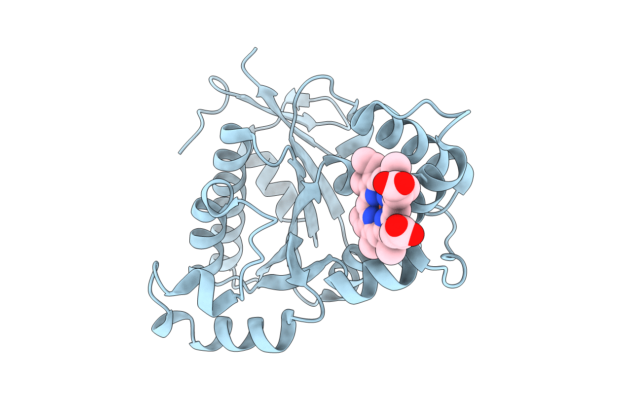
2G5G image
Deposition Date 2006-02-22
Release Date 2006-10-03
Last Version Date 2024-11-13
Entry Detail
PDB ID:
2G5G
Title:
Cofacial heme binding to ChaN of Campylobacter jejuni
Biological Source:
Source Organism(s):
Expression System(s):
Method Details:
Experimental Method:
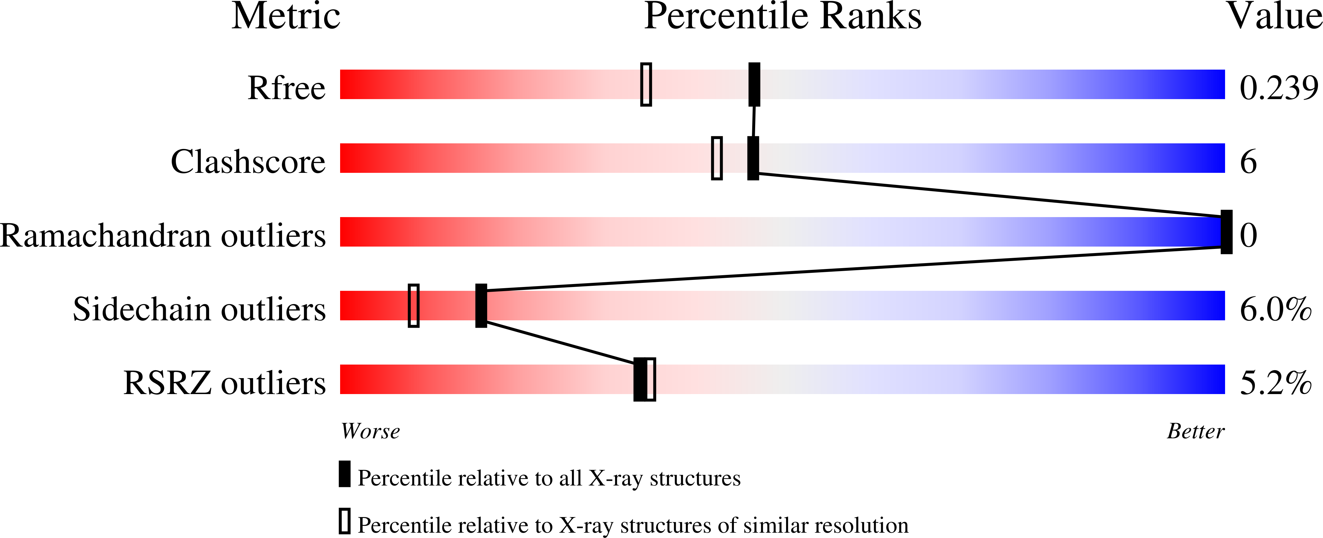
Resolution:
1.90 Å
R-Value Free:
0.24
R-Value Work:
0.20
R-Value Observed:
0.20
Space Group:
C 1 2 1
Macromolecular Entities
Structures with similar UniProt ID
Protein Blast
Polymer Type:polypeptide(L)
Molecule:putative lipoprotein
Chain IDs:A (auth: X)
Chain Length:268
Number of Molecules:1
Biological Source:Campylobacter jejuni subsp. jejuni
Modified Residue
Compound ID Chain ID Parent Comp ID Details 2D Image
MSE A MET SELENOMETHIONINE
Ligand Molecules
Primary Citation
Cofacial Heme Binding is Linked to Dimerization by a Bacterial Heme Transport Protein.
J. Mol. Biol. 362 1108 1119 (2006)
PMID: 16950397 DOI: 10.1016/j.jmb.2006.08.001

Abstact

Campylobacter jejuni is a leading bacterial cause of food-borne illness in the developed world. Like most pathogens, C. jejuni requires iron that must be acquired from the host environment. Although the iron preference of the food-borne pathogen C. jejuni is not established, this organism possesses heme transport systems to acquire iron. ChaN is an iron-regulated lipoprotein from C. jejuni proposed to be associated with ChaR, an outer-membrane receptor. Mutation of PhuW, a ChaN orthologue in Pseudomonas aeruginosa, compromises growth on heme as a sole iron source. The crystal structure of ChaN, determined to 1.9 A resolution reveals that ChaN is comprised of a large parallel beta-sheet with flanking alpha-helices and a smaller domain consisting of alpha-helices. Unexpectedly, two cofacial heme groups (approximately 3.5 A apart with an inter-iron distance of 4.4 A) bind in a pocket formed by a dimer of ChaN monomers. Each heme iron is coordinated by a single tyrosine from one monomer, and the propionate groups are hydrogen bonded by a histidine and a lysine from the other monomer. Sequence analyses reveal that these residues are conserved among ChaN homologues from diverse bacterial origins. Electronic absorption and electron paramagnetic resonance (EPR) spectroscopy are consistent with heme binding through tyrosine coordination by ChaN in solution yielding a high-spin heme iron structure in a pH-dependent equilibrium with a low-spin species. Analytical ultracentrifugation demonstrates that apo-ChaN is predominantly monomeric and that dimerization occurs with heme binding such that the stability constant for dimer formation increases by 60-fold.

Legend

Protein

Chemical

Disease

Primary Citation of related structures
Feedback Form
Name
Email
Institute
Feedback