2CSA image
Deposition Date 2005-05-21
Release Date 2005-05-31
Last Version Date 2024-05-22
Entry Detail
PDB ID:
2CSA
Title:
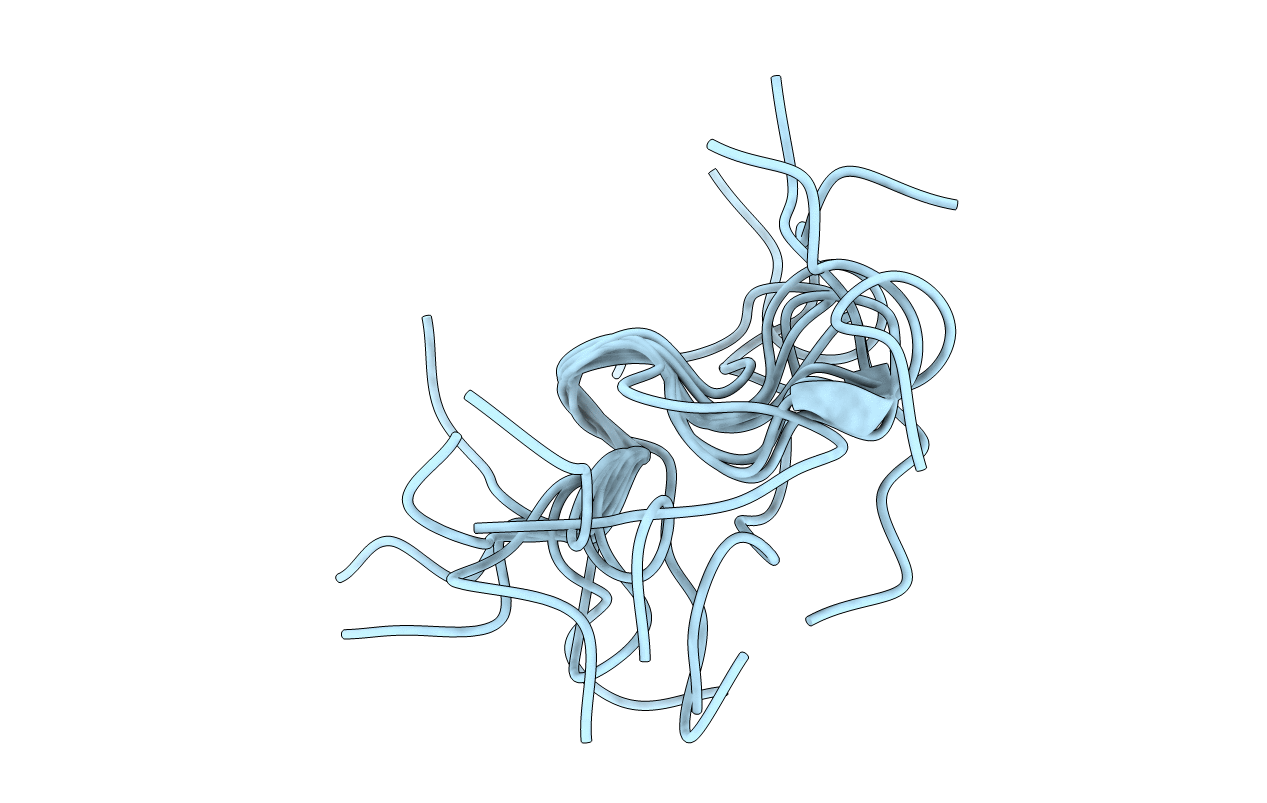
Structure of the M3 Muscarinic Acetylcholine Receptor Basolateral Sorting Signal
Method Details:
Experimental Method:
Conformers Calculated:
200
Conformers Submitted:
10
Selection Criteria:
structures with the lowest energy
Macromolecular Entities
Structures with similar UniProt ID
Protein Blast
Polymer Type:polypeptide(L)
Molecule:Muscarinic acetylcholine rece
Gene (Uniprot):CHRM3
Chain IDs:A
Chain Length:19
Number of Molecules:1
Biological Source:
Ligand Molecules
Primary Citation
Identification and structural determination of the M3 muscarinic acetylcholine receptor basolateral sorting signal.
J. Biol. Chem. 280 24568 24575 (2005)
PMID: 15870063 DOI: 10.1074/jbc.M501264200

Abstact

Muscarinic acetylcholine receptors comprise a family of G-protein-coupled receptors that display differential localization in polarized epithelial cells. We identify a seven-residue sequence, Ala(275)-Val(281), in the third intracellular loop of the M(3) muscarinic receptor that mediates dominant, position-independent basolateral targeting in Madin-Darby canine kidney cells. Mutational analyses identify Glu(276), Phe(280), and Val(281) as critical residues within this sorting motif. Phe(280) and Val(281) comprise a novel dihydrophobic sorting signal as mutations of either residue singly or together with leucine do not disrupt basolateral targeting. Conversely, Glu(276) is required and cannot be substituted with alanine or aspartic acid. A 19-amino acid peptide representing the M(3) sorting signal and surrounding sequence was analyzed via two-dimensional nuclear magnetic resonance spectroscopy. Solution structures show that Glu(276) resides in a type IV beta-turn and the dihydrophobic sequence Phe(280)Val(281) adopts either a type I or IV beta-turn.

Legend

Protein

Chemical

Disease

Primary Citation of related structures
Feedback Form
Name
Email
Institute
Feedback