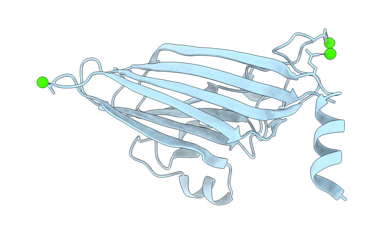
The structure of the protein subunit of satellite tobacco necrosis virus has been solved at 3.7 A resolution. We have now crystallographically refined the original model and extended the resolution ot 2.5 A in order to get a model accurate enough to explain the details of the subunit interactions. The refinement was done with a novel method utilizing the icosahedral symmetry of the virus particle. The final model shows a complicated network of interactions, involving salt linkages, hydrogen bonds and hydrophobic contacts. In addition, we have located three different metal ion sites in the protein shell, linking the protein subunits together. These sites are probably occupied by calcium ions. One site is found in a general position near the icosahedral 3-fold axis of the virus. The ligands form an octahedral arrangement, with two main chain carbonyl oxygens (0-61 and 0-64), one carboxylate oxygen (OD1 from Asp194) of the same subunit and a second carboxylate oxygen (OE1 of Glu25) from a 3-fold related subunit. Two water molecules complete the octahedral arrangement. A second site is on the icosahedral 3-fold axis and is liganded by the carboxylate oxygens of the 3-fold related Asp55 residues. The third metal ion site is found on the 5-fold axis, liganded by the five carbonyl oxygens of Thr138 and two water molecules. We are unable to locate the first 11 N-terminal amino acid residues, which point into the virus interior. No interpretable density for RNA has been found, indicating that the nucleic acid of the virus does not have a unique orientation in the crystal.
Legend
Protein
Chemical
Disease