Deposition Date
2004-07-13
Release Date
2004-07-27
Last Version Date
2023-10-25
Entry Detail
PDB ID:
1U08
Keywords:
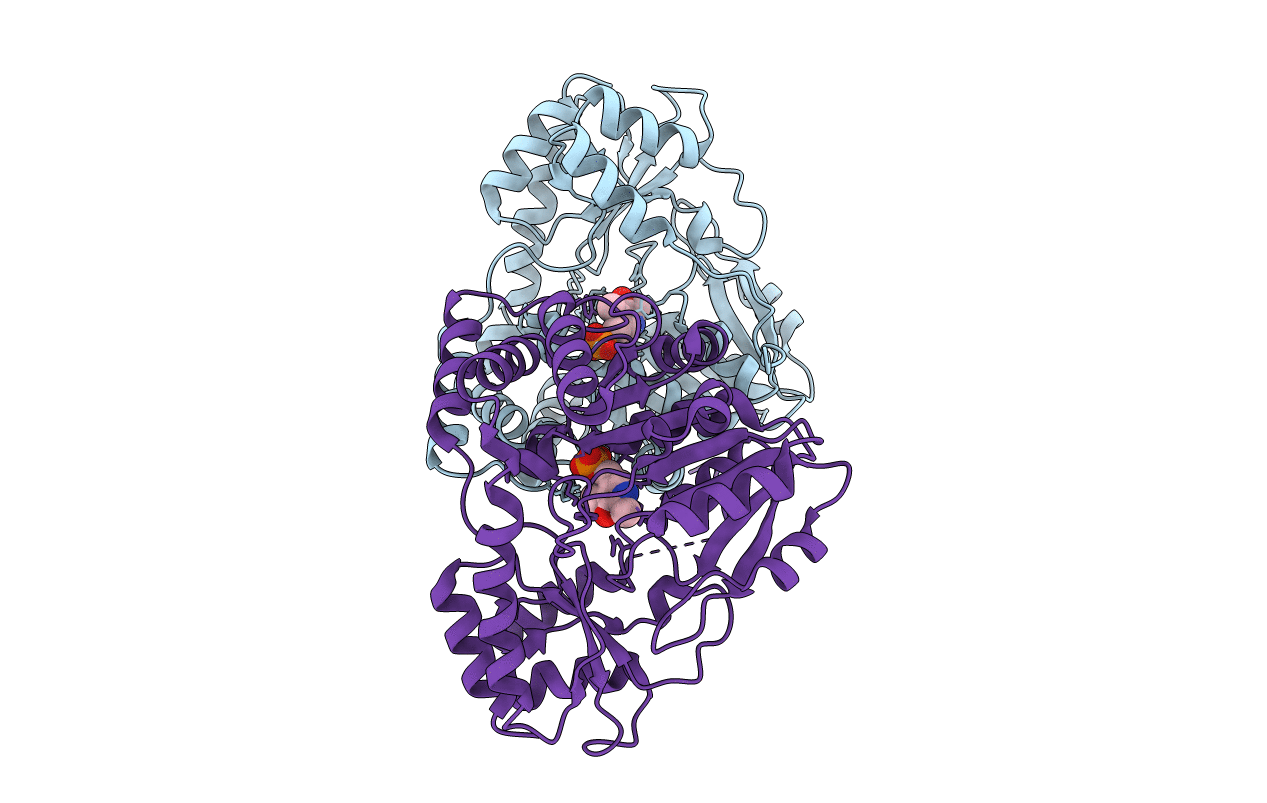
Title:
Crystal Structure and Reactivity of YbdL from Escherichia coli Identify a Methionine Aminotransferase Function.
Biological Source:
Source Organism(s):
Escherichia coli (Taxon ID: 562)
Expression System(s):
Method Details:
Experimental Method:
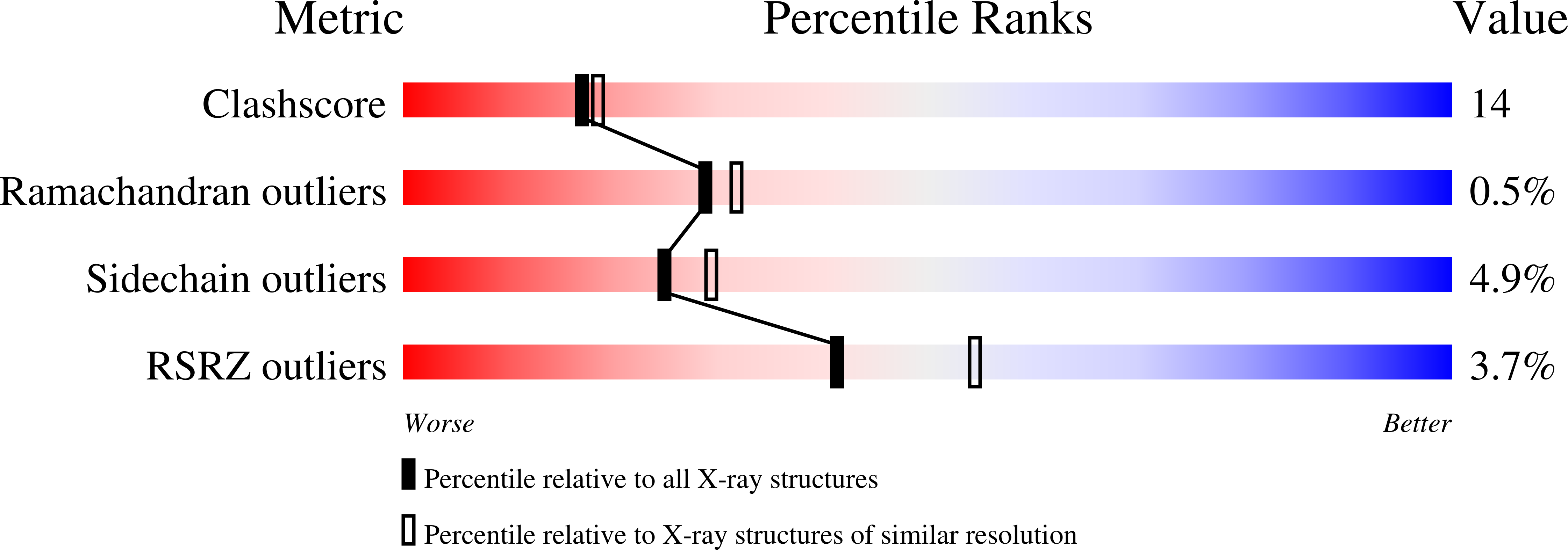
Resolution:
2.35 Å
R-Value Free:
0.22
R-Value Work:
0.19
R-Value Observed:
0.19
Space Group:
P 21 21 21