1QB4 image
Deposition Date 1999-04-30
Release Date 2002-05-01
Last Version Date 2024-02-14
Entry Detail
PDB ID:
1QB4
Keywords:
Title:
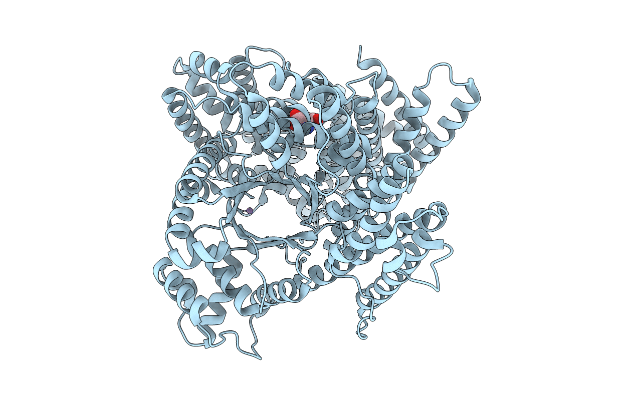
CRYSTAL STRUCTURE OF MN(2+)-BOUND PHOSPHOENOLPYRUVATE CARBOXYLASE
Biological Source:
Source Organism(s):
Escherichia coli (Taxon ID: 562)
Expression System(s):
Method Details:
Experimental Method:
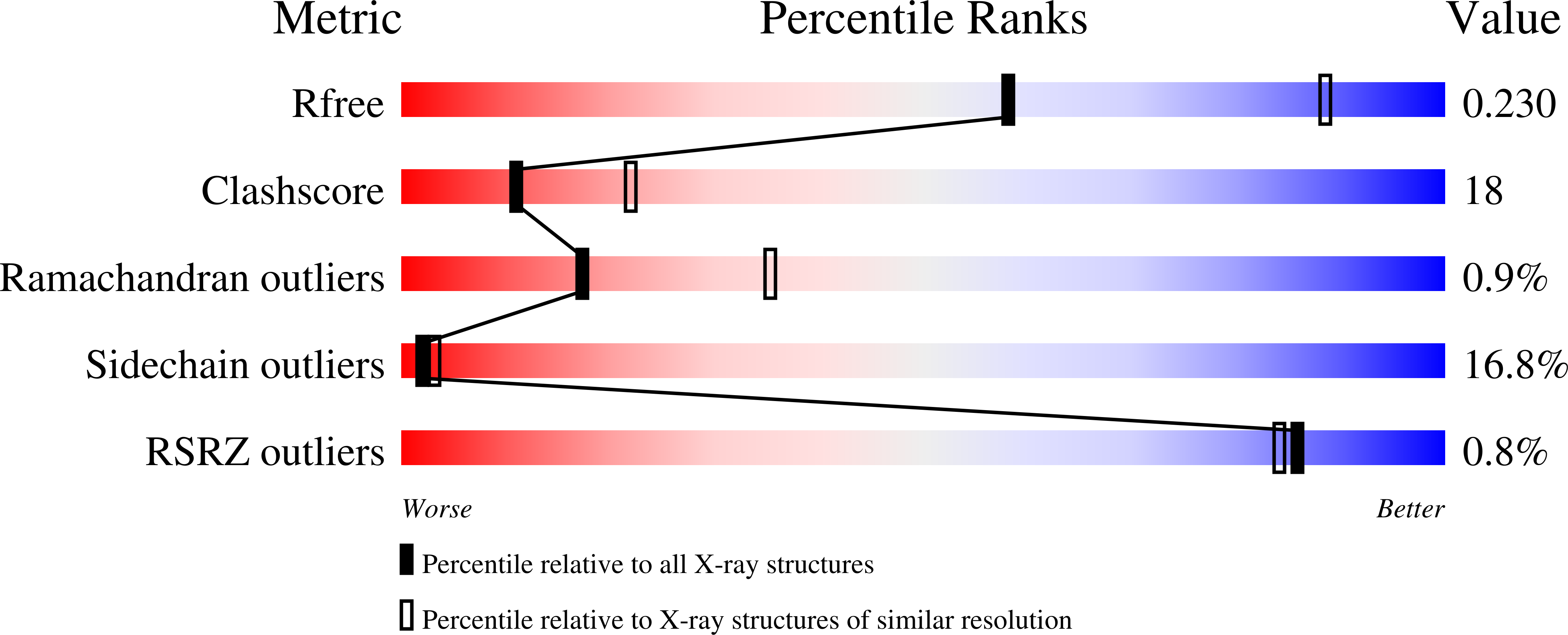
Resolution:
2.60 Å
R-Value Free:
0.26
R-Value Work:
0.22
R-Value Observed:
0.22
Space Group:
I 2 2 2
Macromolecular Entities
Structures with similar UniProt ID
Protein Blast
Polymer Type:polypeptide(L)
Molecule:PHOSPHOENOLPYRUVATE CARBOXYLA
Gene (Uniprot):ppc
Chain IDs:A
Chain Length:883
Number of Molecules:1
Biological Source:Escherichia coli
Primary Citation
Plausible phosphoenolpyruvate binding site revealed by 2.6 A structure of Mn2+-bound phosphoenolpyruvate carboxylase from Escherichia coli
FEBS Lett. 458 93 96 (1999)
PMID: 10481043 DOI: 10.1016/S0014-5793(99)01103-5

Abstact

We have determined the crystal structure of Mn2+-bound Escherichia coli phosphoenolpyruvate carboxylase (PEPC) using X-ray diffraction at 2.6 A resolution, and specified the location of enzyme-bound Mn2+, which is essential for catalytic activity. The electron density map reveals that Mn2+ is bound to the side chain oxygens of Glu-506 and Asp-543, and located at the top of the alpha/beta barrel in PEPC. The coordination sphere of Mn2+ observed in E. coli PEPC is similar to that of Mn2+ found in the pyruvate kinase structure. The model study of Mn2+-bound PEPC complexed with phosphoenolpyruvate (PEP) reveals that the side chains of Arg-396, Arg-581 and Arg-713 could interact with PEP.

Legend

Protein

Chemical

Disease

Primary Citation of related structures
Feedback Form
Name
Email
Institute
Feedback