

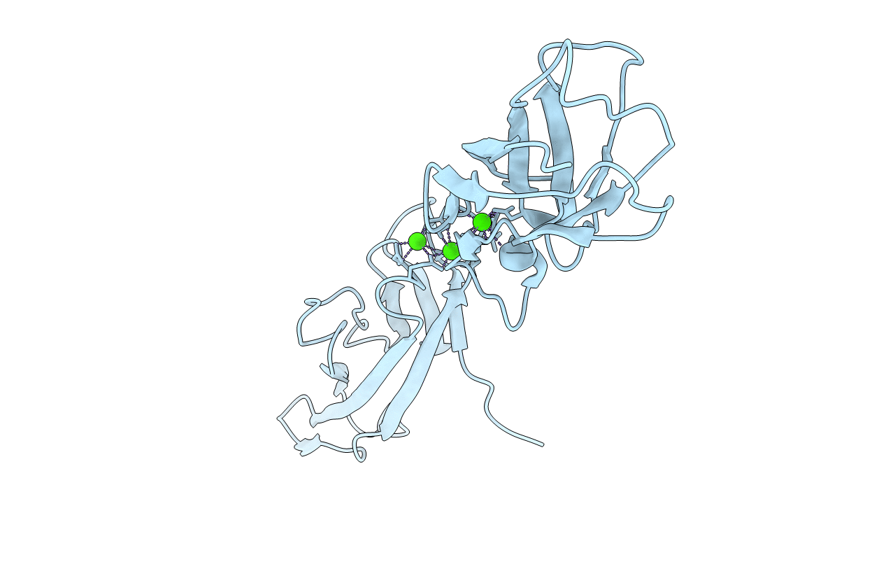
1Q1P image
Deposition Date 2003-07-22
Release Date 2004-04-20
Last Version Date 2023-08-16
Entry Detail
PDB ID:
1Q1P
Keywords:
Title:
E-Cadherin activation
Biological Source:
Source Organism(s):
Mus musculus (Taxon ID: 10090)
Expression System(s):
Method Details:
Experimental Method:
Resolution:
3.20 Å
R-Value Free:
0.30
R-Value Work:
0.25
R-Value Observed:
0.26
Space Group:
C 1 2 1
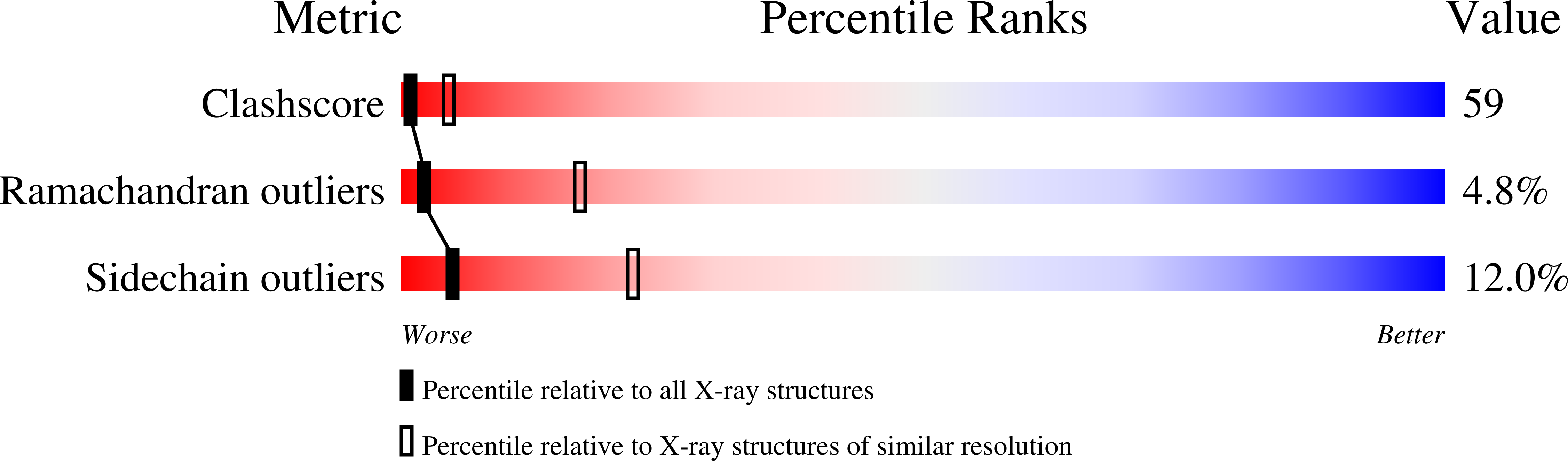
Macromolecular Entities
Structures with similar UniProt ID
Protein Blast
Polymer Type:polypeptide(L)
Molecule:Epithelial-cadherin
Gene (Uniprot):Cdh1
Chain IDs:A
Chain Length:212
Number of Molecules:1
Biological Source:Mus musculus
Ligand Molecules
Primary Citation
Proteolytic E-cadherin activation followed by solution NMR and X-ray crystallography.
EMBO J. 23 1699 1708 (2004)
PMID: 15071499 DOI: 10.1038/sj.emboj.7600192

Abstact

Cellular adhesion by classical cadherins depends critically on the exact proteolytic removal of their N-terminal prosequences. In this combined solution NMR and X-ray crystallographic study, the consequences of propeptide cleavage of an epithelial cadherin construct (domains 1 and 2) were followed at atomic level. At low protein concentration, the N-terminal processing induces docking of the tryptophan-2 side-chain into a binding pocket on the same molecule. At high concentration, cleavage induces dimerization (KD=0.72 mM, k(off)=0.7 s(-1)) and concomitant intermolecular exchange of the betaA-strands and the tryptophan-2 side-chains. Thus, the cleavage represents the switch from a nonadhesive to the functional form of cadherin.

Legend

Protein

Chemical

Disease

Primary Citation of related structures
Feedback Form
Name
Email
Institute
Feedback