1NJJ image
Deposition Date 2002-12-31
Release Date 2003-08-26
Last Version Date 2023-08-16
Entry Detail
PDB ID:
1NJJ
Keywords:
Title:
Crystal structure determination of T. brucei ornithine decarboxylase bound to D-ornithine and to G418
Biological Source:
Source Organism(s):
Expression System(s):
Method Details:
Experimental Method:
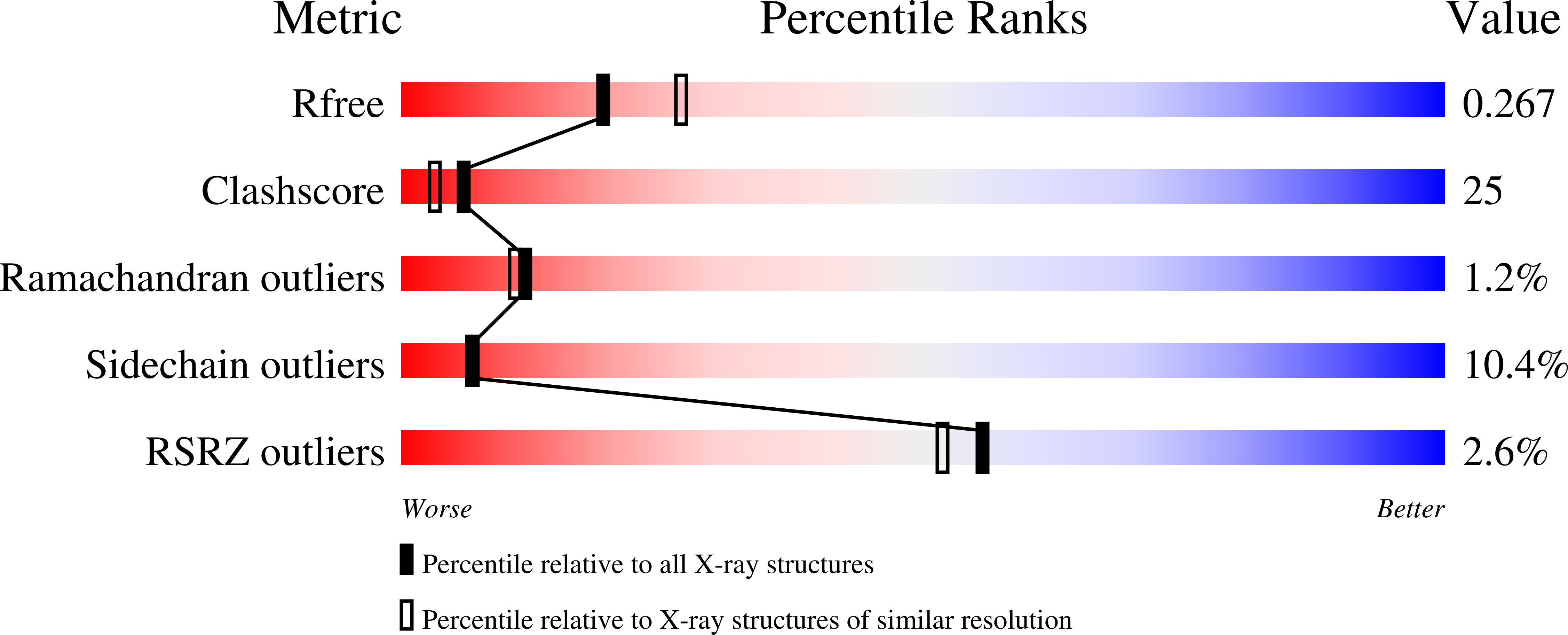
Resolution:
2.45 Å
R-Value Free:
0.28
R-Value Work:
0.26
Space Group:
P 1 21 1
Macromolecular Entities
Structures with similar UniProt ID
Protein Blast
Polymer Type:polypeptide(L)
Molecule:ornithine decarboxylase
Chain IDs:A, B, C, D
Chain Length:425
Number of Molecules:4
Biological Source:Trypanosoma brucei gambiense
Primary Citation
X-ray Structure Determination of Trypanosoma brucei Ornithine Decarboxylase Bound to D-Ornithine and to G418: INSIGHTS INTO SUBSTRATE BINDING AND ODC CONFORMATIONAL FLEXIBILITY.
J.Biol.Chem. 278 22037 22043 (2003)
PMID: 12672797 DOI: 10.1074/jbc.M300188200

Abstact

Ornithine decarboxylase (ODC) is a pyridoxal 5'-phosphate (PLP)-dependent enzyme that catalyzes the rate-determining step in the biosynthesis of polyamines. ODC is a proven drug target to treat African sleeping sickness. The x-ray crystal structure of Trypanosoma brucei ODC in complex with d-ornithine (d-Orn), a substrate analog, and G418 (Geneticin), a weak non-competitive inhibitor, was determined to 2.5-A resolution. d-Orn forms a Schiff base with PLP, and the side chain is in a similar position to that observed for putrescine and alpha-difluoromethylornithine in previous T. brucei ODC structures. The d-Orn carboxylate is positioned on the solvent-exposed side of the active site (si face of PLP), and Gly-199, Gly-362, and His-197 are the only residues within 4.2 A of this moiety. This structure confirms predictions that the carboxylate of d-Orn binds on the si face of PLP, and it supports a model in which the carboxyl group of the substrate l-Orn would be buried on the re face of the cofactor in a pocket that includes Phe-397, Tyr-389, Lys-69 (methylene carbons), and Asp-361. Electron density for G418 was observed at the boundary between the two domains within each ODC monomer. A ten-amino acid loop region (392-401) near the 2-fold axis of the dimer interface, which contributes several residues that form the active site, is disordered in this structure. The disordering of residues in the active site provides a potential mechanism for inhibition by G418 and suggests that allosteric inhibition from this site is feasible.

Legend

Protein

Chemical

Disease

Primary Citation of related structures
Feedback Form
Name
Email
Institute
Feedback