

1NCN image
Deposition Date 2002-12-05
Release Date 2003-03-11
Last Version Date 2024-10-30
Entry Detail
PDB ID:
1NCN
Keywords:
Title:
the receptor-binding domain of human B7-2
Biological Source:
Source Organism(s):
Homo sapiens (Taxon ID: 9606)
Expression System(s):
Method Details:
Experimental Method:
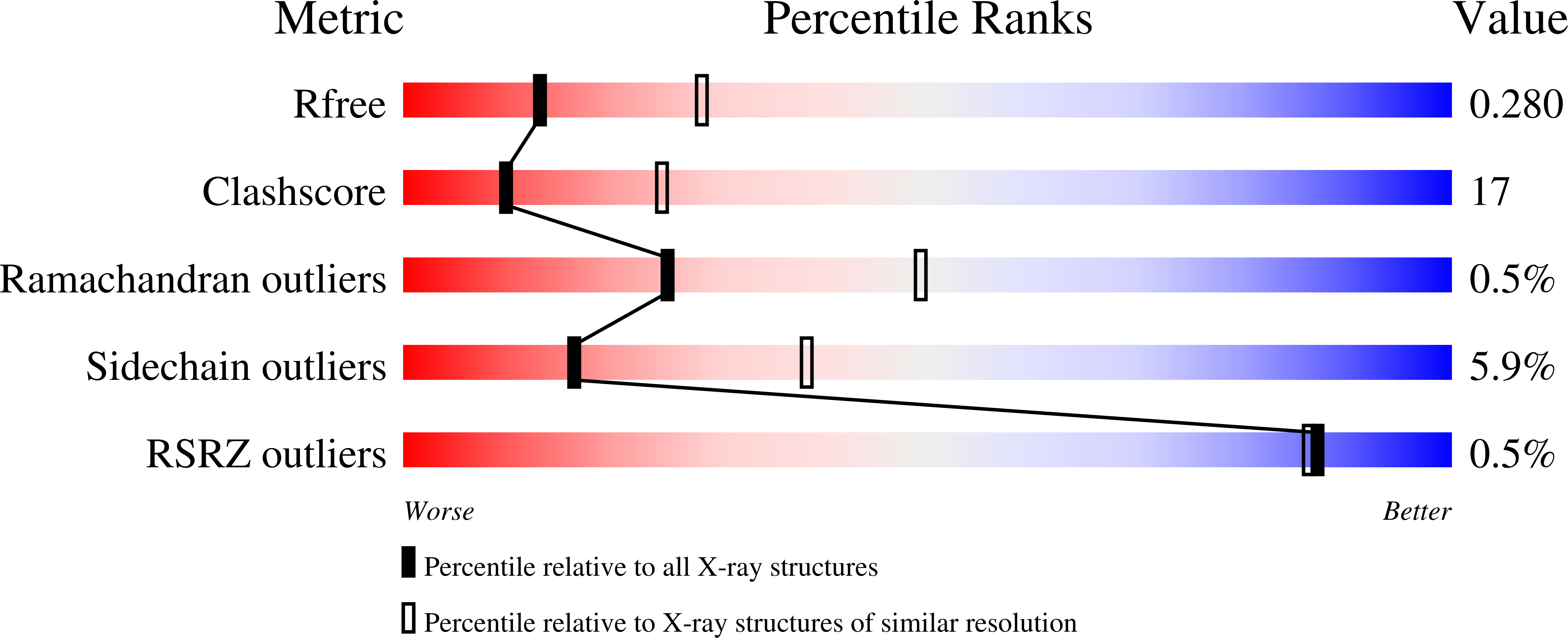
Resolution:
2.70 Å
R-Value Free:
0.27
R-Value Work:
0.21
R-Value Observed:
0.25
Space Group:
P 21 21 2
Macromolecular Entities
Structures with similar UniProt ID
Protein Blast
Polymer Type:polypeptide(L)
Molecule:T lymphocyte activation antig
Gene (Uniprot):CD86
Chain IDs:A, B
Chain Length:110
Number of Molecules:2
Biological Source:Homo sapiens
Ligand Molecules
Primary Citation
Crystal Structure of the Receptor-Binding Domain of Human B7-2: Insights into Organization and Signaling
Proc. Natl. Acad. Sci. U.S.A. 100 2586 2591 (2003)
PMID: 12606712 DOI: 10.1073/pnas.252771499

Abstact

B7-1 and B7-2 are homologous costimulatory ligands expressed on the surfaces of antigen-presenting cells. Their interactions with CD28/CTLA-4 receptors expressed on T cell surfaces are crucial for the proper regulation of T cell activity. B7-1 and B7-2 display distinct roles in immune regulation, although they are usually considered to have redundant functions. Here, we report the crystal structure of the receptor-binding (Ig V-type) domain of human B7-2 at 2.7-A resolution. Structures of unliganded and liganded B7-1 and B7-2 suggest a physical-chemical basis for the observed functional similarities and differences between these two costimulatory ligands. Of particular note, whereas the majority of the residues mediating B7-1 dimerization are hydrophobic, the B7-2 dimer observed in the B7-2/CTLA-4 complex displays a very hydrophilic dimer interface. These differences provide a mechanism for preventing the formation of B7-1/B7-2 heterodimers. The divergence at the putative dimer interface is also consistent with the lower tendency of B7-2 to dimerize, as shown by the monomeric state of unliganded B7-2 both in solution and crystalline form, and may result in detailed differences in signaling mechanisms associated with B7-1 and B7-2.

Legend

Protein

Chemical

Disease

Primary Citation of related structures
Feedback Form
Name
Email
Institute
Feedback