Deposition Date
2002-09-17
Release Date
2002-11-20
Last Version Date
2024-11-13
Entry Detail
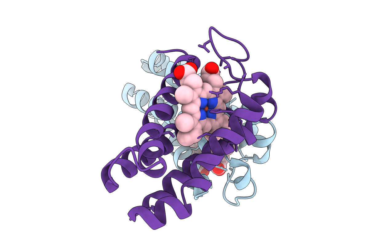
PDB ID:
1MQV
Keywords:
Title:
Crystal Structure of the Q1A/F32W/W72F mutant of Rhodopseudomonas palustris cytochrome c' (prime) expressed in E. coli
Biological Source:
Source Organism(s):
Rhodopseudomonas palustris (Taxon ID: 1076)
Expression System(s):
Method Details:
Experimental Method:
Resolution:
1.78 Å
R-Value Free:
0.26
R-Value Work:
0.21
R-Value Observed:
0.21
Space Group:
P 21 21 21