1LTR image
Deposition Date 1998-07-31
Release Date 1999-02-09
Last Version Date 2024-11-13
Entry Detail
PDB ID:
1LTR
Keywords:
Title:
CRYSTAL STRUCTURE OF THE B SUBUNIT OF HUMAN HEAT-LABILE ENTEROTOXIN FROM E. COLI CARRYING A PEPTIDE WITH ANTI-HSV ACTIVITY
Biological Source:
Source Organism(s):
Escherichia coli (Taxon ID: 562)
Expression System(s):
Method Details:
Experimental Method:
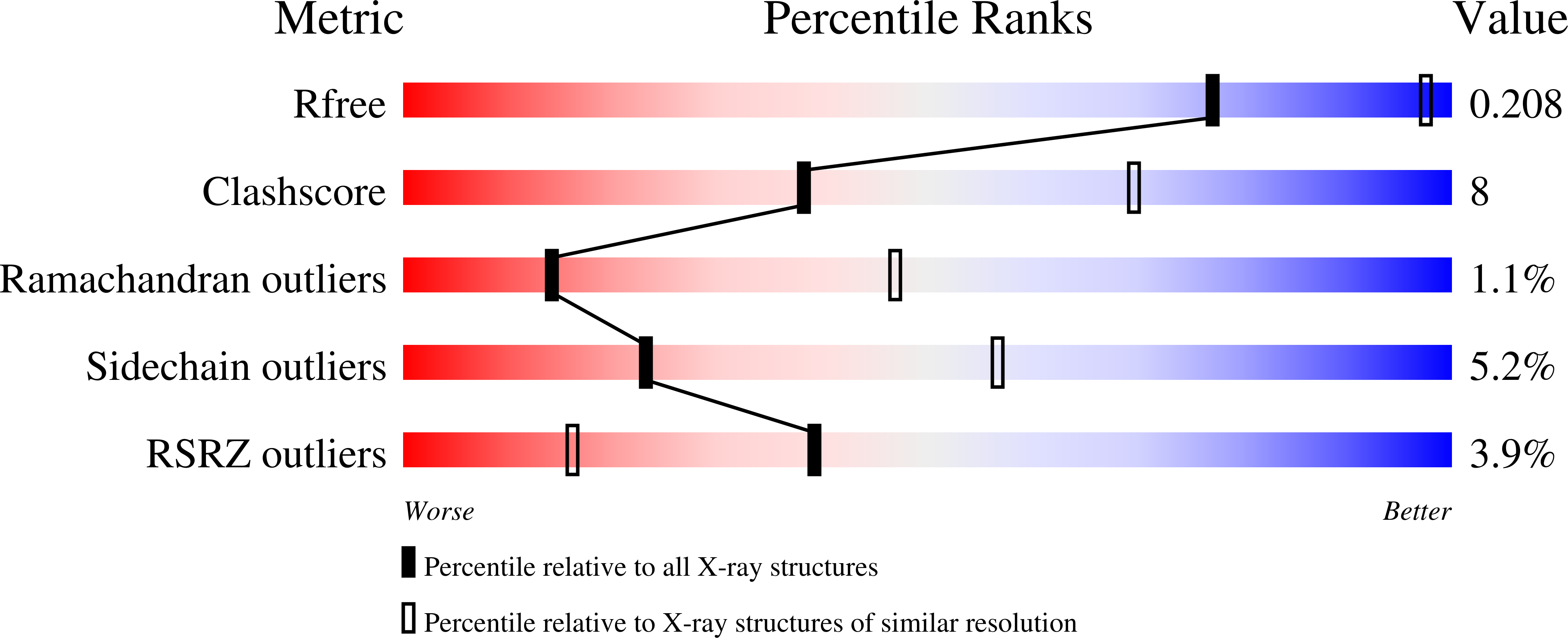
Resolution:
3.04 Å
R-Value Free:
0.21
R-Value Work:
0.18
R-Value Observed:
0.18
Space Group:
P 41 21 2
Macromolecular Entities
Structures with similar UniProt ID
Protein Blast
Polymer Type:polypeptide(L)
Molecule:HEAT-LABILE ENTEROTOXIN
Mutagens:N103K
Chain IDs:A (auth: D), B (auth: E), C (auth: F), D (auth: G), E (auth: H)
Chain Length:113
Number of Molecules:5
Biological Source:Escherichia coli
Ligand Molecules
Primary Citation
Crystal structure of the B subunit of Escherichia coli heat-labile enterotoxin carrying peptides with anti-herpes simplex virus type 1 activity.
J. Biol. Chem. 274 8764 8769 (1999)
PMID: 10085117

Abstact

Two chimeric proteins, consisting of the B subunit of Escherichia coli heat-labile enterotoxin with different peptides fused to the COOH-terminal ends, have been crystallized and their three-dimensional structure determined. The two extensions correspond to (a) a nonapeptide representing the COOH-terminal sequence of the small subunit of herpes simplex virus type 1 ribonucleotide reductase and (b) a 27-amino acid long peptide, corresponding to the COOH-terminal end of the catalytic subunit (POL) of DNA polymerase from the same virus. Both proteins crystallize in the P41212 space group with one pentameric molecule per asymmetric unit, corresponding to a solvent content of about 75%. The overall conformation of the B subunit pentamer in the two chimeric proteins, which consists of five identical polypeptide chains, is very similar to that in the native AB complex and conforms strictly to 5-fold symmetry. On the contrary, the peptide extensions are essentially disordered: in the case of the nonapeptide, only 5 and 6 amino acids were, respectively, positioned in two monomers, while in the other three only 2 residues are ordered. The extension is fully confined to the surface of the pentamer opposite to the face that interacts with the membrane and consequently it does not interfere with the ability of the B subunit to interact with membrane receptors. Moreover, the conformational flexibility of the two peptide extensions could be correlated to their propensity for proteolytic processing and consequent release of a biologically active molecule into cultured cells.

Legend

Protein

Chemical

Disease

Primary Citation of related structures
Feedback Form
Name
Email
Institute
Feedback