1JBA image
Deposition Date 1999-04-03
Release Date 1999-12-10
Last Version Date 2023-12-27
Entry Detail
PDB ID:
1JBA
Keywords:
Title:
UNMYRISTOYLATED GCAP-2 WITH THREE CALCIUM IONS BOUND
Biological Source:
Source Organism(s):
Bos taurus (Taxon ID: 9913)
Expression System(s):
Method Details:
Experimental Method:
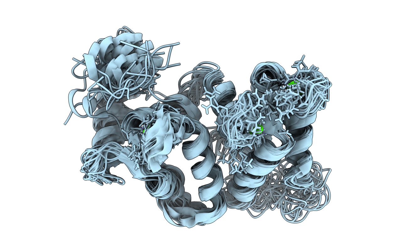
Conformers Calculated:
50
Conformers Submitted:
22
Selection Criteria:
LEAST RESTRAINT VIOLATION
Macromolecular Entities
Structures with similar UniProt ID
Protein Blast
Polymer Type:polypeptide(L)
Molecule:PROTEIN (GUANYLATE CYCLASE AC
Gene (Uniprot):GUCA1B
Chain IDs:A
Chain Length:204
Number of Molecules:1
Biological Source:Bos taurus
Ligand Molecules
Primary Citation
Three-dimensional structure of guanylyl cyclase activating protein-2, a calcium-sensitive modulator of photoreceptor guanylyl cyclases.
J. Biol. Chem. 274 19329 19337 (1999)
PMID: 10383444 DOI: 10.1074/jbc.274.27.19329

Abstact

Guanylyl cyclase activating protein-2 (GCAP-2) is a Ca2+-sensitive regulator of phototransduction in retinal photoreceptor cells. GCAP-2 activates retinal guanylyl cyclases at low Ca2+ concentration (<100 nM) and inhibits them at high Ca2+ (>500 nM). The light-induced lowering of the Ca2+ level from approximately 500 nM in the dark to approximately 50 nM following illumination is known to play a key role in visual recovery and adaptation. We report here the three-dimensional structure of unmyristoylated GCAP-2 with three bound Ca2+ ions as determined by nuclear magnetic resonance spectroscopy of recombinant, isotopically labeled protein. GCAP-2 contains four EF-hand motifs arranged in a compact tandem array like that seen previously in recoverin. The root mean square deviation of the main chain atoms in the EF-hand regions is 2.2 A in comparing the Ca2+-bound structures of GCAP-2 and recoverin. EF-1, as in recoverin, does not bind calcium because it contains a disabling Cys-Pro sequence. GCAP-2 differs from recoverin in that the calcium ion binds to EF-4 in addition to EF-2 and EF-3. A prominent exposed patch of hydrophobic residues formed by EF-1 and EF-2 (Leu24, Trp27, Phe31, Phe45, Phe48, Phe49, Tyr81, Val82, Leu85, and Leu89) may serve as a target-binding site for the transmission of calcium signals to guanylyl cyclase.

Legend

Protein

Chemical

Disease

Primary Citation of related structures
Feedback Form
Name
Email
Institute
Feedback