Deposition Date
2001-05-19
Release Date
2002-02-20
Last Version Date
2024-11-13
Entry Detail
PDB ID:
1J7X
Keywords:
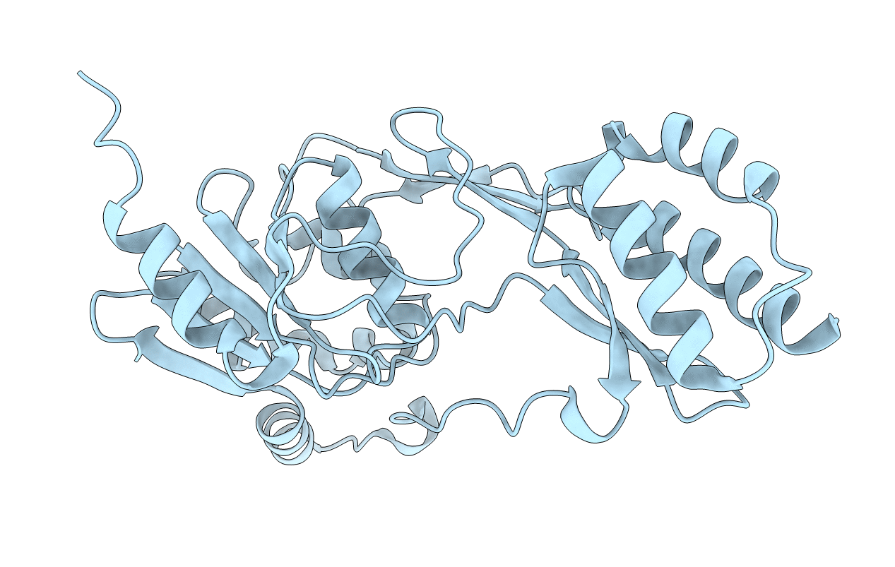
Title:
CRYSTAL STRUCTURE OF A FUNCTIONAL UNIT OF INTERPHOTORECEPTOR RETINOID-BINDING PROTEIN (IRBP)
Biological Source:
Source Organism(s):
Xenopus laevis (Taxon ID: 8355)
Expression System(s):
Method Details:
Experimental Method:
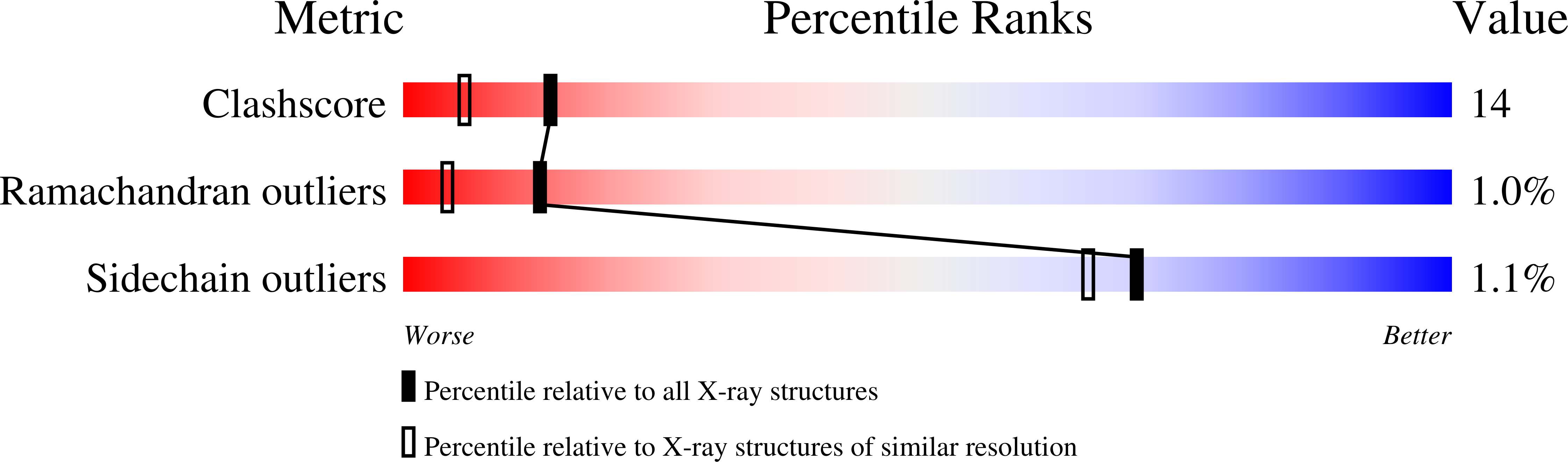
Resolution:
1.80 Å
R-Value Free:
0.22
R-Value Work:
0.20
R-Value Observed:
0.20
Space Group:
P 21 21 21