1H8G image
Deposition Date 2001-02-06
Release Date 2002-01-31
Last Version Date 2024-11-13
Entry Detail
PDB ID:
1H8G
Title:
C-terminal domain of the major autolysin (C-LytA) from Streptococcus pneumoniae
Biological Source:
Source Organism(s):
Expression System(s):
Method Details:
Experimental Method:
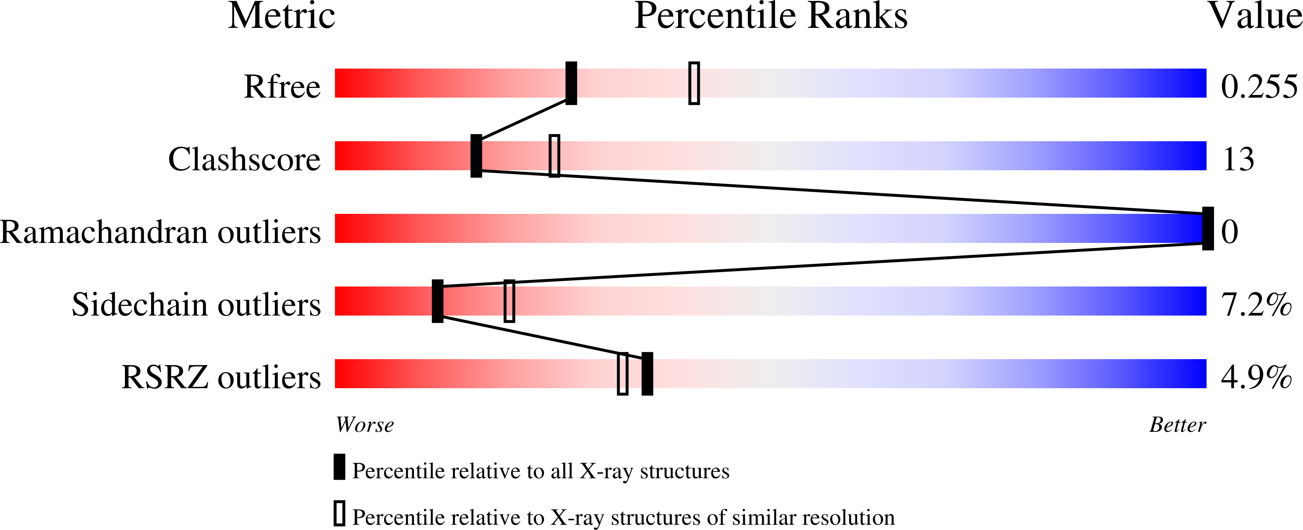
Resolution:
2.40 Å
R-Value Free:
0.26
R-Value Work:
0.21
R-Value Observed:
0.21
Space Group:
P 41 21 2
Macromolecular Entities
Structures with similar UniProt ID
Protein Blast
Polymer Type:polypeptide(L)
Molecule:MAJOR AUTOLYSIN
Gene (Uniprot):lytA
Chain IDs:A, B
Chain Length:95
Number of Molecules:2
Biological Source:STREPTOCOCCUS PNEUMONIAE
Modified Residue
Compound ID Chain ID Parent Comp ID Details 2D Image
MSE A MET SELENOMETHIONINE
Ligand Molecules
Primary Citation
A Novel Solenoid Fold in the Cell Wall Anchoring Domain of the Pneumococcal Virulence Factor Lyta
Nat. Struct. Biol. 8 1020 ? (2001)
PMID: 11694890 DOI: 10.1038/NSB724

Abstact

Choline binding proteins are virulence determinants present in several Gram-positive bacteria. Because anchorage of these proteins to the cell wall through their choline binding domain is essential for bacterial virulence, their release from the cell surface is considered a powerful target for a weapon against these pathogens. The first crystal structure of a choline binding domain, from the toxin-releasing enzyme pneumococcal major autolysin (LytA), reveals a novel solenoid fold consisting exclusively of beta-hairpins that stack to form a left-handed superhelix. This unique structure is maintained by choline molecules at the hydrophobic interface of consecutive hairpins and may be present in other choline binding proteins that share high homology to the repeated motif of the domain.

Legend

Protein

Chemical

Disease

Primary Citation of related structures
Feedback Form
Name
Email
Institute
Feedback