1H5U image
Deposition Date 2001-05-25
Release Date 2001-06-28
Last Version Date 2023-12-13
Entry Detail
PDB ID:
1H5U
Title:
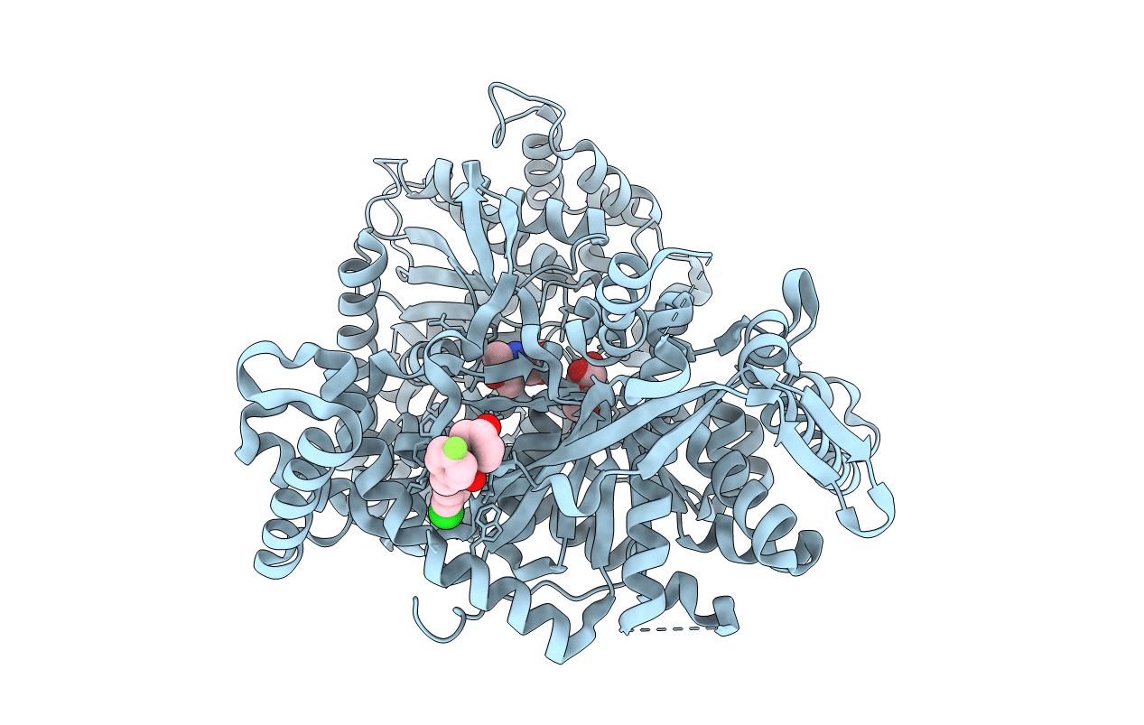
THE 1.76 A RESOLUTION CRYSTAL STRUCTURE OF GLYCOGEN PHOSPHORYLASE B COMPLEXED WITH GLUCOSE AND CP320626, A POTENTIAL ANTIDIABETIC DRUG
Biological Source:
Source Organism(s):
Method Details:
Experimental Method:
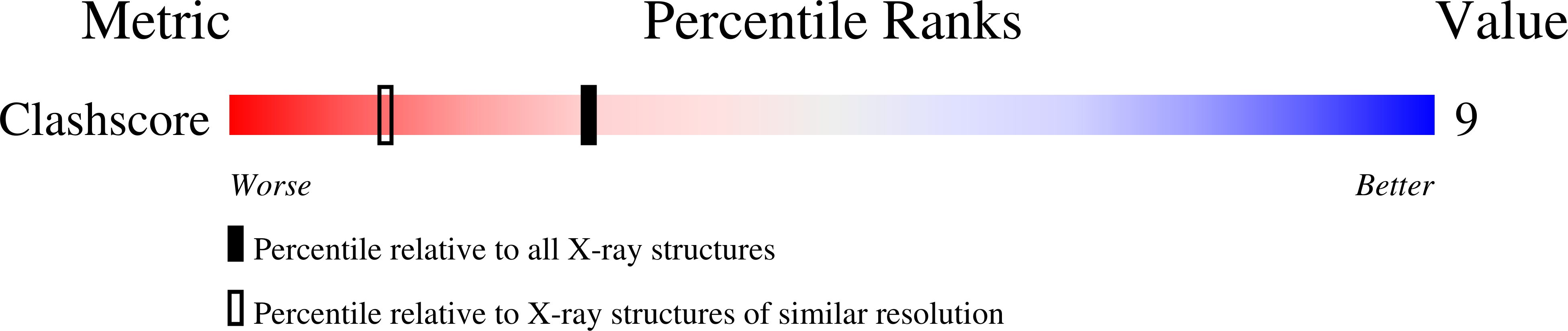
Resolution:
1.76 Å
R-Value Free:
0.23
R-Value Work:
0.21
R-Value Observed:
0.21
Space Group:
P 43 21 2
Macromolecular Entities
Structures with similar UniProt ID
Protein Blast
Polymer Type:polypeptide(L)
Molecule:GLYCOGEN PHOSPHORYLASE
Gene (Uniprot):PYGM
Chain IDs:A
Chain Length:842
Number of Molecules:1
Biological Source:ORYCTOLAGUS CUNICULUS
Primary Citation
The 1.76 A Resolution Crystal Structure of Glycogen Phosphorylase B Complexed with Glucose, and Cp320626, a Potential Antidiabetic Drug
Bioorg. Med. Chem. 10 1313 ? (2002)
PMID: 11886794 DOI: 10.1016/S0968-0896(01)00394-7

Abstact

CP320626, a potential antidiabetic drug, inhibits glycogen phosphorylase in synergism with glucose. To elucidate the structural basis of synergistic inhibition, we determined the structure of muscle glycogen phosphorylase b (MGPb) complexed with both glucose and CP320626 at 1.76 A resolution, and refined to a crystallographic R value of 0.211 (R(free)=0.235). CP320626 binds at a novel allosteric site, which is some 33 A from the catalytic site, where glucose binds. The high resolution structure allows unambiguous definition of the conformation of the 1-acetyl-4-hydroxy-piperidine ring supported by theoretical energy calculations. Both CP320626 and glucose promote the less active T-state, thereby explaining their synergistic inhibition. Structural comparison of MGPb--glucose--CP320626 complex with liver glycogen phosphorylase a (LGPa) complexed with a related compound (CP403700) show that the ligand binding site is conserved in LGPa.

Legend

Protein

Chemical

Disease

Feedback Form
Name
Email
Institute
Feedback