1GPX image
Deposition Date 1998-06-10
Release Date 1999-01-27
Last Version Date 2024-05-01
Entry Detail
PDB ID:
1GPX
Title:
C85S GAPDX, NMR, 20 STRUCTURES
Biological Source:
Source Organism(s):
Expression System(s):
Method Details:
Experimental Method:
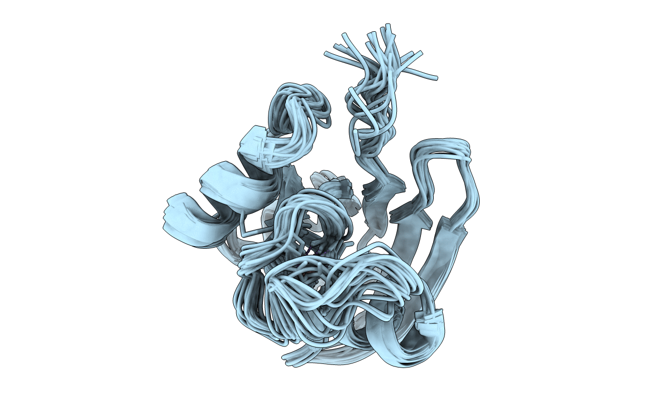
Conformers Calculated:
100
Conformers Submitted:
20
Selection Criteria:
LEAST RESTRAINT VIOLATION
Macromolecular Entities
Structures with similar UniProt ID
Protein Blast
Polymer Type:polypeptide(L)
Molecule:PUTIDAREDOXIN
Gene (Uniprot):camB
Mutagens:C85S
Chain IDs:A
Chain Length:106
Number of Molecules:1
Biological Source:Pseudomonas putida
Ligand Molecules
Primary Citation
The solution structure of a gallium-substituted putidaredoxin mutant: GaPdx C85S.
J.Biomol.NMR 12 407 415 (1998)
PMID: 9835048 DOI: 10.1023/A:1008354113765

Abstact

The Fe2S2 cluster of the ferredoxin putidaredoxin (Pdx) can be replaced by a single gallium ion, giving rise to a colorless, diamagnetic protein in which, apart from the metal binding site, the major structural features of the native ferredoxin are conserved. The solution structure of the C85S variant of gallium putidaredoxin (C85S GaPdx), in which a non-ligand cysteine is replaced by a serine, has been determined via multidimensional NMR methods using uniformly 15N, 13C labeled samples of C85S GaPdx. Stereospecific assignments of leucine and valine methyl resonances were made using 13C, 1H HSQC spectra obtained with fractionally 13C-labeled samples, and backbone dihedral angle restraints were obtained using a combination of two-dimensional J-modulated 15N, 1H HSQC and three-dimensional (HN)CO(CO)NH experiments. A total of 1117 NOE-derived distance restraints were used in the calculations, including 454 short range (i-j < or = 3), 456 long range (i-j > or = 4) interresidue restraints and 207 non-trivial intraresidue restraints. 97 phi and 55 chi 1 angular restraints were also included in the calculation of a family of 20 structures using a combined distance geometry-simulated annealing protocol. Most regions of the protein are well defined in the calculations, with an RMSD of 0.525 A for backbone atoms excluding the metal binding loop (residues 34-48) and the last three C-terminal residues (residues 103-106). Where comparison is possible, these regions show an increase in dynamic behavior over the native protein, as does the loop containing residues 74-76. Structural and dynamic differences between native Pdx and GaPdx are discussed in relation to charge and packing of the metal binding site.

Legend

Protein

Chemical

Disease

Primary Citation of related structures
Feedback Form
Name
Email
Institute
Feedback